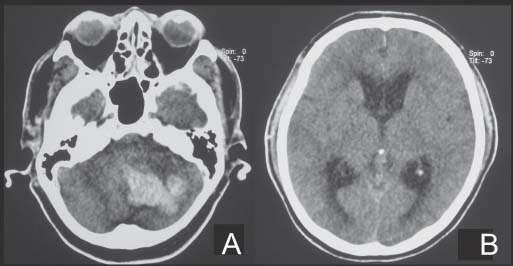
Case 39 Cerebellar Hemorrhage Fig. 39.1 Plain computed tomography scan showing left cerebellar hemorrhage (A) with e ffacement of 4th ventricle and (B) enlargement of the lateral ventricles.
 Clinical Presentation
 Questions
 Answers
< div class='tao-gold-member'>
39 Cerebellar Hemorrhage
Only gold members can continue reading. Log In or Register to continue

Full access? Get Clinical Tree


