45 Chronic Subdural Hematoma

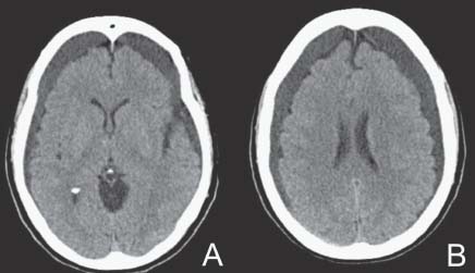
Case 45 Chronic Subdural Hematoma


Remi Nader


Image

Fig. 45.1 (A,B) Computed tomography scan of the brain without contrast.


Image Clinical Presentation


Image Questions


Image

Fig. 45.2 (A) Computed tomography scan with contrast and (B) magnetic resonance imaging fluid-attenuated inversion-recovery axial image without contrast, and T1-weighted images with contrast, (C) axial and (D) coronal.


Image Answers


  1. Interpret the CT scan.

    • CT scan of the brain reveals bilateral subdural hematomas which are mainly chronic with a very small subacute component on the right side.
    • The right subdural hematoma is close to 1.5 cm in thickness. The left subdural hematoma is ~1.3 cm in thickness. They are both mainly along the convexity in the frontal and somewhat parietal area.
    • There is some diffuse brain atrophy in both frontal lobes consistent with advanced Alzheimer’s disease.
    • There also appears to be no midline shift, no obvious mass effect, and no edema. The basal cisterns are wide open.
    • There might be some effacement in the sulci along the cortical surface in the frontal lobes.

  2. What other questions or information would you like to obtain to further plan your management?

    • Obtain current medication history, especially whether or not she is on anticoagulants.
    • Determine if there is a living will or a durable power of attorney. If so, determine the patient’s wishes in a situation where lifesaving measures are needed.

  3. What further studies or investigations would you like to obtain?

    • Laboratory tests: complete blood count, electrolytes, coagulation profile, type and screen
    • Other imaging: MRI – unlikely to change your management in this case
    • If surgical intervention is contemplated, then medical clearance may be needed (electrocardiogram [EKG], chest x-ray, cardiac echography, etc.)

  4. What would you tell the family, next of kin, or caregiver?

  5. How would you approach this case surgically? Describe all the steps of surgery including positioning, incision, bone opening, evacuation, and closure.

    • One operative option consists of bilateral burr hole evacuation of the subdural hematoma, with two burr holes placed on each side.59
    • Preoperative preparation includes

      • Preoperative antibiotics, cefazolin 1 g intravenously (IV) every 8 hours
      • Preoperative seizure prophylaxis with phenytoin, loading dose of 1 g IV over 1 hour, followed by a maintenance dose of 100 mg every 8 hours
      • Positioning is supine with head of bed slightly elevated
      • Anesthesia preferably conscious sedation, or general endotracheal if the patient will tolerate it (i.e., no cardiac or pulmonary risk factors)

    • Surgical steps include

      • Hair clipping and mark incisions along the convexity in-line with each other such that if the need for a bone flap arises it will suffice to connect both incisions.
      • The incisions should be ~5–6 cm away from the midline; one along the parietal boss and one just behind the hair line anteriorly – both ~3 cm in length.
      • Start by opening the right side as this is the symptomatic side.
      • Open skin, galea, and periosteum with a no. 10 blade; expose the skull and perform a burr hole with a pneumatic drill, ~2.5 cm in diameter.
      • Wax the bone edges and cauterize the dura.
      • Open the dura in cruciate fashion, then drain the blood.
      • Irrigate both burr holes profusely with normal saline until the drainage becomes clear.
      • One may place a red rubber catheter in the burr hole subdurally and further irrigate to dislodge any pockets of blood that are harder to access.
      • Closure is completed by placing a piece of Gelfoam (Pfizer, Inc., New York, NY) on top of the burr hole, close the skin and galea with 3–0 Vicryl (Ethicon, Somerville, NJ) and staples.
      • One may leave a small drain in the subgaleal space.

    • The patient is kept with the head of the bed flat postoperatively.10

  6. Preoperatively, as you plan the surgery, she develops hyponatremia with sodium of 118, and she becomes more lethargic. What are some causes of hyponatremia and which is the most likely in this case?

    • Causes of hyponatremia are listed below.
    • Hypovolemic hyponatremia: sodium and free water loss with inappropriately hypotonic fluid replacement

      • Cerebral salt-wasting syndrome: traumatic brain injury, aneurysmal subarachnoid hemorrhage, subdural hematoma, and postcraniotomy – this is the most likely cause in this case
      • Gastrointestinal losses such as vomiting or diarrhea
      • Excessive sweating
      • Third spacing of fluids (peritonitis, pancreatitis, burns)
      • Acute or chronic renal insufficiency
      • Prolonged exercise in a hot environment

    • Euvolemic hyponatremia: normal sodium stores and total body excess free water

      • Psychogenic polydipsia
      • Hypotonic intravenous (i.v.) or irrigation fluids postoperatively

    • Hypervolemic hyponatremia: inappropriate increase in sodium stores

      • Hepatic cirrhosis
      • Congestive heart failure
      • Nephrotic syndrome
      • Hypothyroidism
      • Cortisol deficiency
      • Syndrome of inappropriate antidiuretic hormone (SIADH)11
      • Medications: acetazolamide, thiazide diuretics, angiotensin-converting enzyme inhibitors, carbamazepine, gabapentin, haloperidol, heparin, ketorolac, loop diuretics, nimodipine, opiates, proton pump inhibitors, selective serotonin reuptake inhibitors1113

  7. How would you manage this problem?

  8. What is your course of action now?

    • Urgent ICU admission
    • Laboratory studies: complete blood count, electrolytes, coagulation profile, type and screen, blood, urine, sputum cultures, and culture discharge from wound
    • Obtain imaging studies: CT and MRI with and without contrast
    • May elect to wait until the cultures are sent intra-operatively before starting any broad spectrum antibiotics.
    • Antibiotics suggested:

      • Vancomycin 1 g i.v. every 12 hours, cefepime 2 g i.v. every 12 hours, and metronidazole 500 mg i.v. every 8 hours
      • Gentamycin 600 mg i.v. every 8 hours may be used instead of cefepime if the patient is allergic to penicillin.
      • Antibiotic changes are performed as culture and sensitivity results are obtained.

  9. A follow-up CT scan and subsequently an MRI are obtained (Fig. 45.2). Interpret the studies and provide a differential diagnosis.

    • CT scan with contrast shows some reaccumulation of fluid bilaterally. The fluid collection on the right side is partly hyperdense or enhancing, which is suggestive of acute blood or possible empyema with peripheral enhancement.
    • On MRI, the subdural fluid is consistent with blood of different ages and it is seen bilaterally. On the right side there is some enhancement surrounding the subdural fluid which is possibly suggestive of infection.
    • The right subdural fluid appears to be iso- to hyperintense on T1-weighted and hyperintense on T2-weighted or fluid-attenuated inversion-recovery (FLAIR) MRIs, which, if it is blood, is likely subacute to chronic (3 weeks old).
    • The differential diagnosis includes

      • Reaccumulation of subdural hematoma
      • Subdural empyema
      • Subdural hygroma

  10. What is your management plan?

    • Urgent surgical evacuation via burr hole reexploration on the right side.1416
    • The exposure should be prepared in the event that a craniotomy is needed.
    • Subdural fluid needs to be sent for culture as soon as possible, after which the antibiotics are initiated.
    • Incision and sharp debridement of the posterior scalp wound is needed with irrigation and antibiotics and saline solution.
    • A drain can be left in the subgaleal space.
    • Wound closure can be done in a single layer with 2–0 Prolene (Ethicon, Somerville, NJ) vertical mattress interrupted sutures to promote better healing of infected or granulating tissues.
< div class='tao-gold-member'>

Only gold members can continue reading. Log In or Register to continue

Stay updated, free articles. Join our Telegram channel

Jul 16, 2016 | Posted by in NEUROSURGERY | Comments Off on 45 Chronic Subdural Hematoma

Full access? Get Clinical Tree

Get Clinical Tree app for offline access