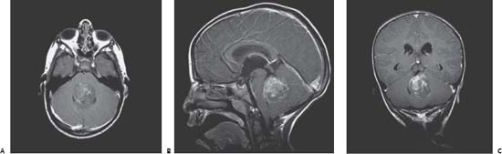
57 Cerebellar Medulloblastoma

Case 57 Cerebellar Medulloblastoma


Ali Raja, Ian F. Pollack, and Nazer H. Qureshi


Image Clinical Presentation


Image

Fig. 57.1 T1-weighted magnetic resonance image (with gadolinium contrast) of the brain. (A) Axial, (B) sagittal, and (C) coronal views showing a large, contrast-enhancing 4th ventricular tumor.


Image Questions


  1. What is your differential diagnosis based on clinical and imaging findings?
  2. What other imaging study would you recommend?
  3. Where do medulloblastomas arise and what are the different histopathologic subtypes?
  4. What is the definitive management of this lesion? What is the role of surgery?
  5. What surgical approaches can be used for resection of this tumor?
  6. Is adjuvant treatment indicated, and if so, what would you recommend?
  7. If you recommend radiation, what is the dose of radiation you would use?
  8. How often is ventriculoperitoneal shunt required in patients with medulloblastomas? What can be a shuntrelated complication in these patients?
  9. Describe a grading system used that affects prognosis in medulloblastoma patients.

Image Answers


  1. What is your differential diagnosis based on clinical and imaging findings?


  2. What other imaging study would you recommend?


    • MRI of the entire spine with and without contrast is also indicated to evaluate for drop metastases or “sugar-coating” along the spinal cerebrospinal fluid (CSF) pathways.

  3. Where do medulloblastomas arise and what are the different histopathologic subtypes?


    • The origin of medulloblastoma is from the roof of the 4th ventricle.
    • The World Health Organization (WHO) classification divides medulloblastomas into four histopathologic subtypes2:


      • Classic medulloblastoma
      • Desmoplastic medulloblastoma
      • Large cell medulloblastoma
      • Medulloblastomas with extensive nodularity

  4. What is the definitive management of this lesion? What is the role of surgery?


    • Maximal surgical resection along with chemotherapy and craniospinal axis radiation would be the indicated treatment.
    • Gross total resection, when possible, can reduce the required radiation dose, with the caveat that the operative goal is not complete microscopic resection (which is the case for other tumors like ependymomas).

  5. What surgical approaches can be used for resection of this tumor?


    • The surgical approach for resection of this tumor can be transvermian, transcortical, or telovelo-cerebellar, among others.3
    • Preoperative corticosteroids may help decrease peritumoral edema with any approach.
    • In cases with ventricular dilatation, insertion of a ventriculostomy in the operating room, via either a coronal or occipital route, immediately prior to the tumor resection, can assist in achieving brain relaxation during the resection and managing CSF diversion following the resection.

  6. Is adjuvant treatment indicated, and if so, what would you recommend?


    • The adjunct therapy would include chemotherapy (various regimens using cisplatin, vincristine, lomustine, and cyclophosphamide are commonly used) along with radiation.

  7. If you recommend radiation, what is the dose of radiation you would use?


    • Standard fractionated radiation regimen for medulloblastomas consists of ~3600 cGy to the entire craniospinal axis with an 1800 cGy boost to the tumor bed.4
    • The so-called average – or standard – risk medulloblastomas (e.g., typical histology, extensively resected, nonmetastatic [M0], posterior fossa lesions in children older than 3 years) have a significantly higher 5-year progression-free survival (60–80%) with this treatment than high-risk tumors (e.g., anaplastic histology, extensive residual disease, metastases, or nonposterior fossa tumor location, and those diagnosed in children younger than 3 years) for which it is less than 40%.4,5
    • The neuraxis dose may be reduced to 2340 cGy with adjuvant chemotherapy in the average-risk group.5
    • Accordingly, reduced-dose radiation is usually not pursued in patients with high-risk tumors, and current studies are examining ways to improve long-term survival rates.
    • Reduced doses of craniospinal radiation for the average risk group may thus diminish cognitive sequelae, although the extent to which doses can be safely reduced is currently undergoing study.
    • Enrollment of patients on multiinstitutional studies to address such issues is essential to ensure that patients receive state-of-the-art postsurgical therapy, which is constantly evolving, and to contribute to improvements in the management of these tumors.

  8. How often is ventriculoperitoneal shunt required in patients with medulloblastomas? What can be a shuntrelated complication in these patients?


    • Less than half the patients require permanent CSF shunting.1
    • Shunt-related intraperitoneal tumor spread is a possible complication, but is rare.6

  9. Describe a grading system used that affects prognosis in medulloblastoma patients.


    • The Chang system (Table 57.1) may be used for evaluating tumor grade in individual patients.
    • For prognostic factors, please refer to Case 21 (Posterior Fossa Tumor).
< div class='tao-gold-member'>

Only gold members can continue reading. Log In or Register to continue

Stay updated, free articles. Join our Telegram channel

Jul 16, 2016 | Posted by in NEUROSURGERY | Comments Off on 57 Cerebellar Medulloblastoma

Full access? Get Clinical Tree

Get Clinical Tree app for offline access