64 Frontal Abscess with Sinus Involvement

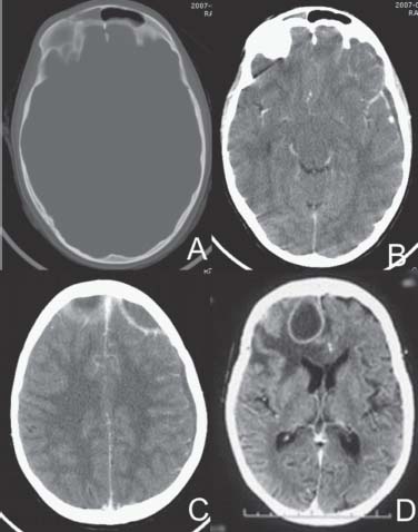
Case 64 Frontal Abscess with Sinus Involvement


Ramez Malak and Rober t Moumdjian


Image

Fig. 64.1 Computed tomography scan of the brain with (A) bone windows and (B–D) brain windows contrast enhanced.


Image Clinical Presentation


Image Questions


  1. Describe the CT scan.
  2. Give a differential diagnosis.
  3. Describe etiologies of cerebral abscess.
  4. Provide predisposing conditions for cerebral abscess.
  5. What is your initial management?
  6. What are some possible complications?
  7. Provide poor prognostic factors.
  8. Describe treatment options and their indications.
  9. Outline follow-up.

Image Answers


  1. Describe the CT scan.

    • There is a ring-enhancing lesion in the left frontal subdural space and fullness of the right frontal sinus.

  2. Give a differential diagnosis.

  3. Describe etiologies of cerebral abscess.

    • Between 30 and 60% of pyogenic abscesses are mixed infections, with aerobic isolates outnumbering anaerobic isolates ~2 to 1.
    • Organisms include streptococci, Staphylococcus aureus, and gram-negative bacilli.
    • In neutropenic patients, brain abscesses may be caused by Candida or Aspergillus.
    • In immunosuppressed patients, the causes may include Toxoplasma gondii, mycobacterium, Nocardia, Cryptococcus, or Listeria.

  4. Provide predisposing conditions for cerebral abscess.

    • Immunosuppression, diabetes mellitus, steroid use, alcoholism, renal failure, intravenous drug abuse, meningitis (especially in children, 12% of pediatric abscess), sinusitis, mastoiditis, postoperative craniotomy, ear, nose, and throat surgery or dental surgery, cyanotic heart disease in children (tetralogy of Fallot)
    • Systemic infection

      • Skin pustules, folliculitis, pulmonary infections, osteomyelitis, dental abscess, and subacute bacterial endocarditis

    • Right-to-left shunting
    • Lung arteriovenous fistula, Osler–Weber–Rendu syndrome, patent foramen ovale

      • Removal of organisms from the systemic circulation by the lungs is bypassed.2,3

  5. What is your initial management?

    • Admission
    • Septic workup: blood culture, chest radiography, urine culture, blood count
    • Sedimentation rate and C-reactive protein
    • Cardiac echography
    • Drainage of air sinus or mastoids
    • Lumbar puncture is contraindicated.
    • Antimicrobial therapy

      • If no causative pathogen is identified, initial empirical antimicrobial therapies are selected in accordance with the portal of entry and the anatomic location of the abscess (Table 64.1). For example, penicillin G, metronidazole, and third-generation cephalosporins are initially indicated for brain abscess associated with sinusitis.

    • Once the organism is identified, change antibiotics according to sensitivity.
    • Duration of parenteral antimicrobial therapy is 6 to 8 weeks provided that the etiologic organisms are susceptible and that adequate surgical drainage can be obtained. It is recommended that antibiotics are continued until complete disappearance of the enhancement of the capsule.
    • Corticosteroids

      • May be beneficial in patients with increased ICP from edema and potentially life-threatening complications, provided they are given after antibiotic therapy is started. However, they could delay immune responses and encapsulation and may decrease enhancement of the abscess wall on CT.

    • Anticonvulsant therapy

      • Seizures are frequent complications of brain abscess. We recommend that seizure prophylaxis or antiepileptic medication be given in every case and continued for extended periods.

  6. What are some possible complications?

    • Vascular thrombosis
    • Brain infarctions
    • Ventriculitis (requires ventricular drainage and intraventricular and systemic antibiotics)
    • Hydrocephalus
    • Empyema of epidural or subdural spaces
    • Subdural effusions
    • Recurrence after aspiration (3–25%), after excision (0–6%)3
    • Death
    • Long-term sequelae: seizures, focal neurologic deficits, and cognitive dysfunction

  7. Provide poor prognostic factors.

  8. Describe treatment options and their indications.

    • Medical treatment

      • Nonsurgical, empirical treatment is possible and efficient in certain patients, especially when the etiologic agent is known, abscess is less than 2 cm, or at the stage of cerebritis.

    • Surgical treatment

      • Burr hole drainage, simple needle aspiration, or stereotactic aspiration

        • Advantages: simple; it can be used in the cerebritis stage; has less potential morbidity than surgical excision, especially in case of edema
        • Indications: Abscesses in the cerebritis stages, deep-seated abscesses, multiple abscesses, abscesses located in eloquent areas of the brain, abscesses extending in various sinuses and mastoids, subdural empyema
        • Risks: rupture of the abscess into the ventricle or leakage into the subarachnoid space, intracerebral hematoma
        • Contraindications: multiloculated abscesses


      • Excision

        • Advantages: lower incidence of recurrence, shorter hospitalization
        • Indications: posterior fossa abscesses, multiloculated abscesses, failure of multiple aspirations, adhesions to the dura, abscesses caused by more resistant pathogens such as fungi, abscesses containing gas, posttraumatic abscesses containing foreign bodies or contaminated retained bone fragments, abscesses resulting from fistulous communication (dermal sinus)
        • Disadvantages: higher morbidity, increased brain edema
        • Contraindications: abscesses in the cerebritis stages, deep-seated abscesses in eloquent areas, multiple abscesses

  9. Outline follow-up.

    • The clinical improvement can precede radiologic improvement. Complete resolution of the abscess cavity will take ~12 weeks. Residual contrast enhancement should not dictate the need for additional therapy. Usually intravenous antibiotics are administered for 6 weeks, generally followed by oral intake for 3 weeks.
    • The effectiveness of the treatment of these patients should be judged according to clinical status and neuroradiographic findings. CT should show decreases in degree of ring enhancement, edema, mass effect, and size of the lesion.
    • Regular follow-up by CT should be at least every 3 months to document the therapeutic response and complete resolution of the ring-enhancing abscesses.1,3
< div class='tao-gold-member'>

Only gold members can continue reading. Log In or Register to continue

Stay updated, free articles. Join our Telegram channel

Jul 16, 2016 | Posted by in NEUROSURGERY | Comments Off on 64 Frontal Abscess with Sinus Involvement

Full access? Get Clinical Tree

Get Clinical Tree app for offline access