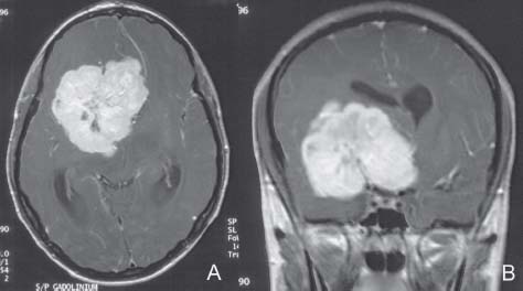
8 Hemangiopericytoma

Case 8 Hemangiopericytoma


Burak Sade and Joung H. Lee


Image

Fig. 8.1 (A) T1-weighted postcontrast axial and (B) coronal magnetic resonance images showing a diffusely enhancing 8-cm perisellar mass originating from the anterior clinoid process. Note the significant midline shift and ventricular dilatation.


Image Clinical Presentation


Image Questions


  1. What are the significant findings in the MRI?
  2. What is your differential diagnosis?
  3. What would be your initial management?
    The patient was operated through a right frontotemporal craniotomy. The surgery had to be staged because of extensive bleeding. Gross total resection was achieved after the second stage (
    Fig. 8.2). The tumor histology confirmed the diagnosis of hemangiopericytoma.
  4. How is hemangiopericytoma classified in the most recent World Health Organization (WHO) classification in 2007, and how does it differ from meningioma in this classification?
  5. Is there any role for adjuvant treatment?
    Following surgery, the patient received intensity modulated radiotherapy (IMRT).
        Seven years after his initial surgery and radiotherapy, he had a recurrence to the left ventral petrous region, which was treated with surgery and IMRT. Nine years after his initial surgery, he developed recurrence to his original tumor bed, which was resected.
        At the time of his second recurrence, a metastasis was detected in his pancreas, which was treated with radiotherapy. Currently, he is 11 years out of his first surgery, with no intracranial detectable disease and stable pancreatic metastasis.
  6. What is the prognosis for hemangiopericytoma?
< div class='tao-gold-member'>

Only gold members can continue reading. Log In or Register to continue

Stay updated, free articles. Join our Telegram channel

Jul 16, 2016 | Posted by in NEUROSURGERY | Comments Off on 8 Hemangiopericytoma

Full access? Get Clinical Tree

Get Clinical Tree app for offline access