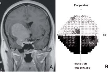
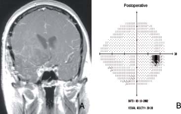
9 Clinoidal Meningioma

Case 9 Clinoidal Meningioma


Burak Sade and Joung H. Lee


Image

Fig. 9.1 (A) T1-weighted postcontrast coronal magnetic resonance image showing a 7 cm enhancing mass arising from the anterior clinoid process and compressing the chiasm laterally and superiorly. (B) Visual field examination is shown on the right and reveals an inferior arcuate visual field defect.


Image

Fig. 9.2 (A) Postoperative T1-weighted postcontrast coronal magnetic resonance image showing gross total resection of the tumor with minimal residual within the cavernous sinus. (B) Postoperative visual field examination is shown on the right and reveals resolution of the defect.


Image Clinical Presentation


Image Questions


  1. What is your differential diagnosis?
  2. How would you classify clinoidal meningiomas?
  3. How would you initially manage this patient?
  4. What are your treatment goals and options?
  5. What other evaluations would you like to have before the surgery?
    Management: The patient was operated on through a right frontotemporal craniotomy using a skull-base approach, which enables early decompression of the optic nerve and enhanced exposure and resectability of the tumor.
    13 Gross total resection was achieved with small residual in the cavernous sinus (Fig. 9.2). The histology was grade I meningothelial meningioma. Her retroorbital headache, visual acuity loss, and field defect resolved completely following surgery (Fig. 9.2).
  6. What are the critical structures in the vicinity of clinoidal meningiomas?
  7. What is your follow-up plan?

Image Answers


  1. What is your differential diagnosis?

    • Based on the location and imaging characteristics of the lesion, differential diagnoses include meningioma and hemangiopericytoma.
    • Other less likely diagnoses include glioma, lymphoma, metastases, or abscess.

  2. How would you classify clinoidal meningiomas?

  3. How would you initially manage this patient?

    • Admit to the ward or intensive care unit.
    • Obtain laboratory studies (complete blood count, electrolytes, prothrombin time, partial thromboplastin time, type and screen)
    • Steroids (dexamethasone). This might provide some relief with her pressure headache and some improvement of her visual deficits.
    • Seizure prophylaxis. The history of “funny smell” is suspicious for temporal lobe seizures. Therefore, seizure prophylaxis would be reasonable.

  4. What are your treatment goals and options?

    • The patient presents with a 7-cm lesion causing significant mass effect and visual deterioration. Therefore, surgery is indicated.
    • The main goals of the surgery would be to decompress the optic nerve and achieve as maximum resection as possible.1 Because of the age of the patient, extent of resection is important as a significant residual will likely result in tumor regrowth in her lifetime.
    • A tumor of this size cannot be treated with gamma-knife stereotactic radiosurgery. However, if she were to have significant medical comorbidities, which would make surgery not feasible, fractionated radiotherapy could be an option.46
    • Conservative management is not advisable in her case.

  5. What other evaluations would you like to have before the surgery?

    • Neuroophthalmologic consultation. In this location, even in patients who do not present with visual symptoms, this assessment is warranted because of the proximity of the tumor to the optic nerve and the frequent involvement of the optic canal by the tumor.
    • Computed tomography (CT) scan of the head to evaluate for bone remodeling and possible hyperostosis4
    • Angiography. The role of angiography is controversial. In selected cases, it may be helpful in demonstrating the critical vascular structures, and it may be used to embolize the tumor preoperatively in some cases.4

  6. What are the critical structures in the vicinity of clinoidal meningiomas?

    • Critical neurovascular structures include the optic and oculomotor nerves, internal carotid, middle cerebral, anterior cerebral arteries and their branches, pituitary stalk, and contents of the cavernous sinus (especially in tumors with cavernous sinus extension).2,3

  7. What is your follow-up plan?

    • The histology of the tumor and extent of resection are the main factors that dictate the follow-up plan.
    • Simpson grading can be helpful in evaluating likelihood of recurrence (see Case 7, Sphenoid Wing Meningioma).
    • In this case, because of the grade I histology and gross total resection, no further treatment is necessary at this point. The patient can be followed up by imaging periodically.
    • In the case of a recurrence, the recurrent portion’s proximity to the optic nerve will dictate the management strategy.4,5
< div class='tao-gold-member'>

Only gold members can continue reading. Log In or Register to continue

Stay updated, free articles. Join our Telegram channel

Jul 16, 2016 | Posted by in NEUROSURGERY | Comments Off on 9 Clinoidal Meningioma

Full access? Get Clinical Tree

Get Clinical Tree app for offline access