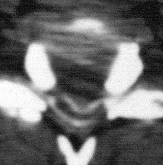
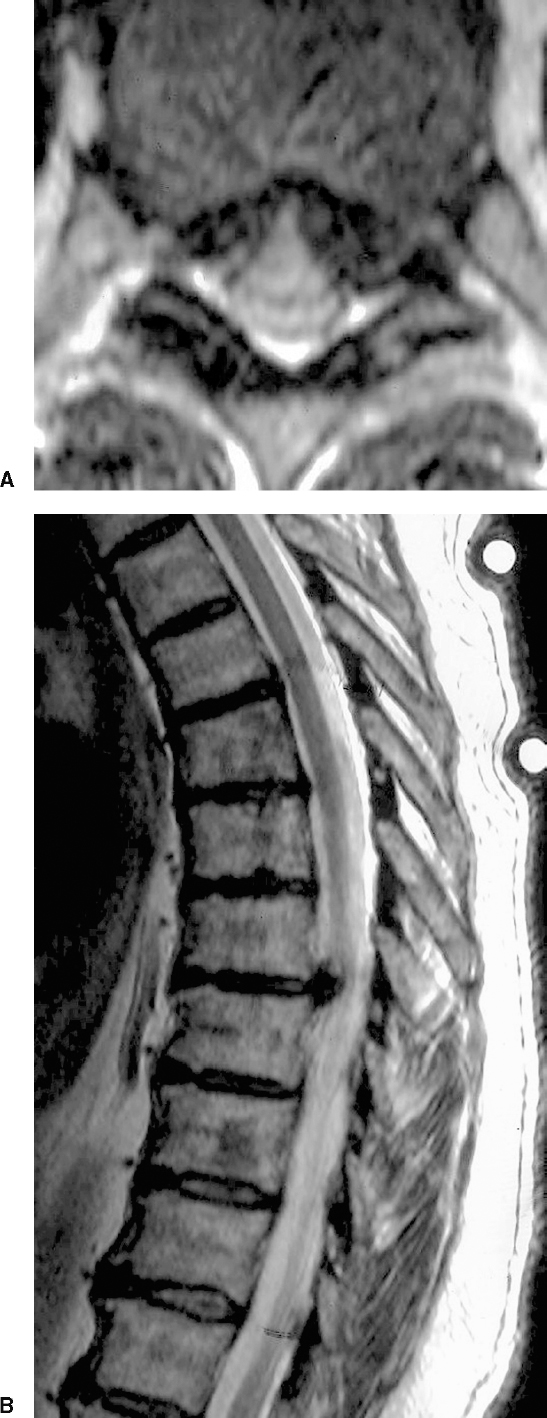
17 Acute Intervention for Cervical, Thoracic, and Lumbar Spinal Disk Disease John B. Pracyk and Vincent C. Traynelis The natural history of spinal disk disease is often benign and self-limited. Symptoms may present acutely or with gradual progression over weeks to months. Patients with persistent sensory disturbances, pain, or mild neurological deficits secondary to disk herniations are initially treated nonoperatively with immobilization, traction, analgesics, and physical therapy, and such cases do not require emergency surgery. Eighty to ninety percent of these individuals obtain symptomatic relief with medical management only.1 Approximately 10% of patients fail to improve despite maximum conservative therapy and are referred for surgical evaluation. Surgical decompression, if indicated, is performed electively in these cases. In contrast, 0.25% to 1.0% of patients with herniated disks present with severe deficits or rapidly progressive neurological deterioration.2,3 These patients may exhibit symptoms and signs that include marked radicular weakness, myelopathy, and bowel or bladder dysfunction. Inappropriate or delayed treatment of these individuals may result in increased morbidity or persistent neurological deficit. This chapter focuses on the role of acute surgical intervention in patients with severe or progressive neurological symptoms secondary to spinal disk disease. Aging predisposes the intervertebral disk to pathological changes. Metabolic transport through the end plate decreases as the disk ages and can lead to abnormalities in macromolecular synthesis and subsequent disk degradation. Desiccation and internal disruption of the nucleus pulposus result in loss of disk volume. These degenerative processes predispose the intervertebral disk to the formation of anulus fibrosus fissures.4,5 Increased intradiskal pressure can result in herniation of the nucleus pulposus through these anular fissures. Disk herniation usually occurs in patients with mild to moderate degenerative changes. Other factors that may increase the risk of disk herniation include trauma, rotational forces, and axial loading. Extreme axial loading with rotation may produce acute herniations in relatively normal intervertebral disks in the lumbar spine. The majority of patients, however, do not report any specific activity preceding the sudden onset of symptoms associated with acute disk herniations. The initial evaluation of patients includes a careful history, which often assists in distinguishing between vascular, infectious, neoplastic, or traumatic causes of acute neurological deterioration. The neurological examination should be recorded in detail and the time of the evaluation documented. This provides a functional baseline for subsequent examinations. Initial radiographic studies, including anteroposterior (AP), lateral, and oblique x-rays, are useful for assessing alignment and bony anatomy. Selected patients may also benefit from information obtained with flexion/extension lateral radiographs. Additional imaging methods are usually necessary to accurately diagnose the etiology of the acute deficit. These include computed tomography (CT), magnetic resonance imaging (MRI), and myelography. Plain radiographs are used to evaluate alignment, stability, bony anatomy, and degenerative disk disease but are inadequate in the diagnosis of acute disk herniation. Indeed, plain films have been reported as “normal” in 20% to 50% of acute herniations.1,6–9 Approximately one third of patients with disk herniations will have disk space narrowing evident on plain films; however, this is a common radiographic finding, especially in individuals over the age of 50 years.10 Calcification of extruded disk fragments may assist in identifying the appropriate interspace; however, this finding is present in only 25% of cases and indicates long-standing pathology, and additional imaging is therefore necessary. Compared with myelography and CT, MRI allows direct imaging of neural structures and provides the greatest soft-tissue detail. Multiplanar, gadolinium-enhanced T1- and unenhanced T1- and T2-weighted images assist in differentiating neurological deterioration secondary to disk herniation, contusion, syrinx, infection, tumor, infarction, or demyelinating or degenerative spinal cord diseases. Numerous additional imaging paradigms enhance the diagnostic capabilities of MRI. Currently, MRI is the single best noninvasive study for diagnosing a herniated disk.11 There are limitations of MRI for visualizing the spine. For example, false-positive images may occur with cerebrospinal fluid (CSF) flow artifact; however, false-negatives are uncommon. In addition, bony anatomy is poorly visualized with MRI; however, compression from coexisting osteophytes can be inferred from thecal sac indentation or spinal cord deformity. Thin-slice, multiplanar T1- and T2-weighted images reduce partial volume averaging, CSF flow void artifact, and signal drop-out from calcified herniated disks, thereby improving visualization of the spinal cord and nerve roots.12,13 CT provides greater detail of the bony structures as compared with MRI, but soft-tissue definition is usually inadequate to satisfactorily determine the extent of neural compression. Additionally, large disk fragments in the lumbar spine that occupy the canal may easily be overlooked.14 Although the diagnostic sensitivity of CT for disk herniation is ~80%,15 CT without intrathecal contrast is impractical as a screening procedure. Postmyelography CT provides excellent detail of the bony anatomy and neural compression. High-resolution postmyelography CT scanning is significantly more sensitive than myelography alone in diagnosing extreme lateral disk herniations.16 In general, postmyelography CT provides an excellent adjunctive study to further delineate specific anatomy, such as the lateral recess, or to reconcile against an MRI study in which the etiology of clinical symptoms is not demonstrated. Prior to MRI, myelography was the gold standard for imaging herniated disks; however, today it is rarely performed as a “stand-alone” test (i.e., without postmyelography CT).8 Myelography does not provide direct information on the etiology of the lesion or extent of neural compression. Compression of neural structures is inferred by filling defects in the intrathecal contrast. Disadvantages of myelography include decreased diagnostic sensitivity with small herniations and possible complications because it is an invasive technique. Additionally, myelography is usually nondiagnostic in cases of far lateral disk herniation in which nerve root compression is distal to the dural nerve sheath, although the postmyelogram CT can be quite useful in this particular situation. The timing of surgical intervention for symptomatic disk disease is somewhat controversial. Emergent surgical intervention should be reserved for those patients with severe or rapidly progressive motor radiculopathy, myelopathy, or bowel or bladder dysfunction secondary to acute disk herniations. In contrast, patients without evidence of spinal instability who present with pain, sensory disturbances, and mild or fixed motor deficits or those exhibiting neurological improvement should not be considered for emergent surgical decompression. Instead, these patients should be treated with conservative and supportive medical management. If they fail these treatment modalities, then elective surgical intervention should be considered. In 1934, Mixter and Barr reported 19 cases of surgically treated disk herniations producing neural compression.17 Cervical diskectomies accounted for only 4% of disk surgery during the next 20 years following this report.17,18 Inadequate radiographic techniques and the belief that small disk herniations were incapable of producing significant symptoms or neurological deficits frequently resulted in radicular symptoms being misdiagnosed as brachial plexus neuralgia or Spillane’s neuritis, whereas myelopathy was often felt to be secondary to disseminated sclerosis. Improved imaging techniques and a better understanding of disk disease have increased the frequency of diagnosis of symptomatic herniated cervical disks. By 1980, cervical diskectomies accounted for approximately one third of all disk surgeries.8,18 The most commonly affected levels in decreasing order of frequency are C5-C6, C6-C7, and C4-C5. Ninety-five percent of cervical disk herniations occur at these three levels.8 There is a slight male predominance, and the peak age is in the fourth and fifth decades. The risk of developing symptomatic cervical disk disease increases with congenital or degenerative narrowing of the spinal canal.19 Disk herniations may also occur in conjunction with fracture/dislocations or facet dislocations. The symptoms and signs of disk herniation are produced by compression and vascular compromise of the nerve roots or spinal cord. Patients with symptomatic cervical spondylosis may present with radiculopathy, myelopathy, or a combination of both. Acute cervical radiculopathy usually results from lateral or posterolateral disk herniations. Early degenerative changes may produce mild foraminal narrowing secondary to osteophyte formation or facet hypertrophy. These changes can tether or stretch the nerve root so that even relatively small disk herniations may result in profound neurological deficits.18 The exact pathophysiology of radiculopathy remains unclear; however, it appears that both compression as well as ischemia of the nerve root are required to produce symptoms. The entire process may be amplified by the presence of inflammatory mediators. Although pain and sensory abnormalities are the most common complaints of acute cervical disk herniations, -60% of patients will exhibit weakness and hyporeflexia by the time of evaluation.8 Figure 17-1 Axial magnetic resonance image of a C7-T1 disk herniation producing significant spinal cord compression. The patient also has congenital spinal stenosis. This individual presented with acute quadriparesis. Compression of the cervical spinal cord by central and centrolateral disk herniations may result in quadriparesis, painless sensory disturbances, and hyperreflexia (Fig. 17-1). Myelopathy secondary to acute disk herniation is probably the result of both spinal cord compression and vascular compromise, although sudden extrusion of the intervertebral disk may produce neurological deterioration secondary to direct spinal cord pressure or contusion or both.20 In the absence of trauma, the acute onset of neurological deficit is usually due to vascular compromise. Long transverse perforating arteries arising from the anterior spinal artery supply the ventral gray matter as well as the lateral funiculi of the spinal cord.21 Compression of the cord in a ventrodorsal direction compromises these transverse arteries and results in ischemia of the anterior gray matter and lateral white matter tracts. This ischemia produces lower motor neuron signs at the level of compression from anterior horn cell involvement and upper motor neuron findings caudal to the disk herniation secondary to lateral corticospinal tract dysfunction. Patients with evidence of cervical trauma deserve special consideration, especially those with unilateral or bilateral facet dislocations. Facet dislocations with a concomitant traumatic disk herniation can produce cervical cord compression at the level of facet dislocation22–25; however, determining the significance of the disk herniation remains more difficult. Eismont et al reported a series of 63 patients managed with closed traction reduction.26 Open reduction was performed if the closed method failed. Interestingly, only one patient in this series worsened following posterior open reduction and fusion. More importantly, critical examination of these and other cases demonstrated that no awake patient experienced neurological deterioration as a result of a closed reduction procedure.24 The widespread use of MRI in cervical trauma has resulted in the demonstration of disrupted or herniated disks in approximately one third to half of patients with facet dislocations. Not surprisingly, this has prompted many to recommend a prereduction MRI for patients with cervical dislocations. Harrington et al reported a series of 37 patients managed with closed reduction.27 They achieved a 97% rate of successful reduction without neurological morbidity. None of the treated patients developed a permanent neurological deficit as a result of attempted closed reduction, and of those patients who underwent successful closed reduction, none deteriorated. The Joint Section on Disorders of the Spine and Peripheral Nerves of the American Association of Neurological Surgeons and the Congress of Neurological Surgeons has established Guidelines for the Management of Acute Cervical Spine and Spinal Cord Injuries. According to these evidence-based guidelines, several large clinical series have failed to establish a relationship between the presence of a prereduction herniated disk and the development of neurological deterioration with attempted closed traction reduction in awake patients.28 In summary, although prereduction MRI will demonstrate disk herniations in up to half of patients with facet subluxations, the clinical significance of these herniations is debatable; thus the utility of a prereduction MRI in awake patients who can cooperate with a neurological examination is minimal.28 MRI does, however, have a proven role in two specific subsets of patients: patients with cervical spine fracture/dislocations who cannot be examined during attempted closed reduction, and those who require an open reduction. The presence of a significant disk herniation in this setting is a relative indication for ventral decompression. To date, no prospective comparative study of closed reduction versus anterior decompression and stabilization for patients with MRI-documented herniated disks in association with unreduced cervical fracture/dislocation injuries has been performed.28 Historically, posterior and posterolateral approaches were the most frequently used techniques during the first 20 years of cervical disk surgery. A midline incision with lateral subperiosteal dissection exposes the laminae and facet joints. With this approach, a hemilaminotomy and medial facetectomy are required to obtain adequate exposure of the lateral disk space and lateral recess of the spinal canal. Paramedian and lateral disk herniations are more readily accessible than central herniations with a posterior approach.29,30 The risk of postoperative instability is minimal if less than one third of the facet joint is resected. The posterior approach lends itself well to the current trend toward more minimally invasive approaches, with the procedure being performed through an access tube or port.31 This minimizes the surgical incision, and the subperiosteal muscle dissection is replaced with a muscle-splitting approach. Central disk herniations approached posteriorly require extensive bone and facet joint removal for adequate ventral exposure, which increases the risk of postoperative instability. This factor, combined with the potential for spinal cord manipulation, makes this an unfavorable option, particularly with the ease of ventral approaches. Although rarely used today, lateral approaches should be included for a historical perspective, and the anterior lateral approach may be an alternative to posterior facetectomy or anterior diskectomy for herniated cervical disks in highly selected patients.32–35 The skin incision follows the anterior border of the sternocleidomastoid, and soft-tissue dissection is continued until the transverse processes are identified. This approach requires skeletonizing the vertebral artery and retracting it laterally to gain access to the neural foramen. The advantages include direct visualization of the nerve root as it exits the foramen and preservation of the posterior apophyseal joints as well as the supporting ligaments. The disadvantages include the risk of injury to the vertebral artery and sympathetic chain and limited access to the contralateral neural foramen. Anterior cervical diskectomy has been used for -40 years to treat ventral spinal lesions.36 This technique allows access to the entire anterior spinal canal and both neural foramina at each vertebral level. Today, intervertebral body fusion with allograft or autograft with or without anterior cervical plating is often performed upon the completion of the neural decompression. Postoperative neurological outcome is related to the type, duration, acuteness, and severity of the preoperative deficit. Radicular symptoms are more likely to improve with surgical decompression compared with myelopathy; however, several small reports note significant improvement in myelopathic patients if surgery is performed early.30,37–40 There does not seem to be a consensus as to a critical period of time in which further delay produces irreversible deficits. Patients with deficits from acute disk herniations have a more favorable surgical outcome compared with those with deficits from spondylotic disease.8,41 Patients who present with severe or long-standing symptoms and signs have a poorer functional outcome than those with only a short clinical history and minor neurological deficits. Acute disk herniations in the thoracic spine are relatively uncommon compared with the cervical or lumbar regions. The incidence of symptomatic thoracic disk herniations is reported as 1/million/year, and this disease accounts for only 0.2% to 1.5% of all diskectomies.15,42–47 There is a slight male predominance, and most patients are affected in the third through fifth decades.15,43,46,47 Disk herniations have been reported at every level in the thoracic spine; however, 70% to 80% occur below T8. This increased frequency is thought to be secondary to greater mobility at these lower levels.13,15,43,46,47 Multiple thoracic disk herniations are uncommon.42 Herniations occur most often in the midline (>70%) followed by centrolateral and lateral prolapses (Fig. 17-2).42,43,46–48 Associated risk factors for acute disk herniations include lifting or bending, trauma, and Scheuermann’s disease.15 The signs and symptoms of an acute thoracic herniated disk can be divided into radicular presentation or myelopathic presentation. Myeloradiculopathy refers to patients with spinal cord and root signs or symptoms. In general, a laterally displaced herniated disk is more likely to produce radicular symptoms rather than myelopathy. Bandlike pain and sensory abnormalities involving the thorax and abdomen are the most common presentations. These symptoms are frequently misdiagnosed as pleuritis, angina, or cholecystitis. Radicular motor deficits involving the T1 nerve root may result in interosseous wasting and hand weakness. Central or centrolateral disk herniations may produce myelopathy secondary to spinal cord compression and ischemia. The cross-sectional area of the thoracic spinal cord occupies a relatively larger portion of the spinal canal compared with the cervical region; consequently, a small disk herniation can produce a disproportionately significant canal compromise with a resultant myelopathy. Some of the pathophysiology of thoracic spinal cord dysfunction is felt to be related to vascular compromise.45 Central disk herniations present with lower extremity weakness as the initial symptom in 20% to 30% of patients, and more than 50% of patients will have a frank myelopathy at the time of clinical evaluation.13,15,43,44 Bladder and bowel dysfunction are present in 30% to 70% of patients at the time of presentation.13,43,44,48,49 The natural history of symptomatic herniated thoracic disks is usually one of progressive neurological deterioration, usually over several years.
Pathogenesis
Initial Evaluation
Radiographic Evaluation
Indications for Acute Surgical Intervention
Cervical Spine
Clinical Presentation
Cervical Trauma
Surgical Approaches
Posterior
Lateral
Anterior
Prognosis
Thoracic Spine
Clinical Presentation
Acute Intervention for Cervical, Thoracic, and Lumbar Spinal Disk Disease
Only gold members can continue reading. Log In or Register to continue


Full access? Get Clinical Tree


