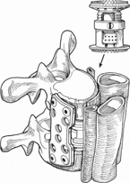
33 John T. Street and Marcel F. S. Dvorak Application of a low profile anterior/anterolateral plating system to the thoracic and thoracolumbar spine. Anterior thoracic and thoracolumbar plating systems provide additional stability by means of neutralization and load-sharing capacity when combined with reconstruction of the anterior and middle columns of the spine. The primary function of anterior plating is to maintain the alignment of a short segment of the thoracic and thoracolumbar spine following direct anterior decompression of the neural elements. Secondary benefits include the stability provided to anterior interbody grafts, improved rates of surgical arthrodesis, minimizing the number of instrumented motion segments, and the avoidance of the need to augment with posterior instrumentation techniques. Thoracic and thoracolumbar segmental instability secondary to anterior and middle column incompetence. This instability is primarily the result of either complete diskectomy or corpectomy for fracture, tumor, infection, degeneration, iatrogenic injury, and failed previous stabilization surgery (either anterior or posterior) resulting in pseudarthrosis. The absolute contraindication to anterior plating techniques relates to the inability to obtain secure fixation of the implants in the adjacent vertebral bodies. This may be due to either disease involvement (infection, tumor) of adjacent vertebrae or to severe osteoporosis. Osteoporosis is known to disproportionately affect the cancellous bone of the vertebral bodies, and knowing this, the surgeon is advised to rely more on what cortical bone is preserved. In osteoporosis, posterior hook and wire fixation is preferred, whereas multisegment disease involvement by tumor or infection is optimally stabilized by adding posterior segmental fixation. Anterior plating techniques, functioning primarily as neutralization devices, require an intact posterior tension band (posterior spinal elements) and thus massive disruption of posterior elements by virtue of injury (flexion distraction fractures) or disease (tumor) make this technique less than ideal. The other major limitation of anterior plate fixation is the limited ability to correct deformity through this approach. Translational and rotational deformity cannot be corrected through anterior vertebral body manipulation, and thus these deformities are contraindications to this approach. Newly acquired kyphotic deformities, where there is “plasticity” in the posterior elements can be corrected through an anterior corpectomy, or release followed by the application of pure distraction through the anterior column. Stiff or long-standing kyphotic deformities require combined approaches and are not amenable to anterior plating alone. Relative contraindications include generalized or local osteopenia, pulmonary function prohibiting thoracotomy, and high risk of not being able to be weaned from the ventilator. Detailed preoperative computed tomography (CT) and magnetic resonance imaging (MRI) is required for planning the surgical approach, decompression, reconstruction, and fixation. Preemptive estimation of plate size and bicortical screw lengths aids intraoperative decision making. Appreciation of three-dimensional anatomy, particularly that of the great vessels, is important. We have seen cases where plain radiographic images are initially interpreted to be vertebral osteomyelitis, which upon closer inspection of the CT scan images are clearly a mycotic aneurysm that has eroded into the vertebral bodies. Preoperative angiography may be indicated to identify the artery of Adamkiewicz. Thoracotomy allows access from T3 to T11. Right-sided thoracotomy is recommended due to the position of the aorta and the azygous vein. Rib resection should be performed one or two levels above the lesion. An extensile cervicothoracic approach should be considered for lesions cephalad to T5 (beware of the thoracic duct on left side and of the recurrent laryngeal nerve on the right). The 10th rib (Dwyer) thoracoabdominal approach, usually performed on the left side to avoid the liver, facilitates exposure caudal to T11 and is extensile down to the L5 vertebral body. Surgery at the incorrect level is a major concern, often underestimated, in all anterior thoracic surgery. Advanced imaging such as CT and MRI often does not aid in the intraoperative determination of the correct level, where poor-quality intraoperative plain films do not facilitate rib visualization or counting up or down the spine. Plain anteroposterior (AP) and lateral scout films on CT or plain radiographs performed preoperatively are mandatory to determine the number of rib-bearing vertebrae, the number of lumbar vertebrae, and the levels of interest. Some techniques that have been used in particularly complex situations (e.g., the obese patient, or obscure anatomy) involve the placement of a needle under the skin, or contrast material in the vertebral body (similar to a vertebroplasty) at the desired operative level in the radiology suite prior to coming to the operating room. Double-lumen endobronchial intubation is required for thoracotomies to allow selective lung deflation. For thoracoabdominal approaches, the lung can simply be retracted and single lung ventilation is not required. Frequent, intermittent, intraoperative lung expansion reduces postoperative atelectasis. Intercostal nerve blocks should be administered under direct vision prior to any rib resection. Neurophysiologic monitoring is highly recommended for cases in which segmental arteries are to be sacrificed and decompressions performed. Ensure that the patient is positioned in as true a lateral position as possible, to aid spatial orientation intraoperatively. An axillary roll should be used. The table may be flexed at the required thoracotomy level to aid access. When the patient is positioned, and prior to draping, an AP and cross-table lateral x-ray should be taken to aid in level identification and orientation. At this time the skin incision level corresponding to the required vertebral level should be clearly marked. These measures will help maintain orientation, optimize screw and plate position, and prevent iatrogenic segmental sagittal and coronal malalignment. During the approach, the segmental arteries can be temporarily clamped prior to ligation to determine if there is any alteration in motor evoked potential (MEP) or somatosensory evoked potential (SSEP) signals. The rib head inserts into the superior part of the corresponding vertebra and extends ventrally to obscure a significant portion of the dorsal disk space. The key to an adequate decompression is to resect the rib head, identify the neural foramen, and then proceed with decompression. Furthermore, resection of the rib heads facilitates true lateral positioning of the plate and screws. There is an inherent tendency to drift anteriorly and place the instrumentation in an anterolateral position, where it is more likely to contact the aorta. This is particularly risky in the more elderly patient with a tortuous aorta, which if it is adjacent to even smooth metal contours, can erode over time as it pulsates against a screw or plate. This has been known to cause fatal exsanguinations to occur in the early and late postoperative time frame. When rodscrew anterior fixation systems are used, great care must be exercised in cutting the rods and positioning them to avoid any sharp burrs or edges on the implants that may come in contact with vital intrathoracic structures. Bone graft may be obtained from the resected vertebral body or from the rib resected in the approach. This bone is often placed anteriorly under the anterior longitudinal ligament. In this position, it is visible on follow-up lateral radiographs as it consolidates into a fusion mass. The pleura may then be closed over the bone graft, plates, and screws to prevent intrathoracic migration of bits of bone graft. The most difficult screws to insert are often the screws in the most cephalad vertebra. The orientation of the end plate (following completion of the decompression) guides the screw trajectory and helps determine the screw length. In the presence of tumor, infection, or previous surgery, safe exposure of sufficient vertebral body may be challenging. If amenable, segmental vessels should be tied as well as clipped in case of inadvertent clip loss. During decompression and dissection in the neural foramen, Gelfoam, Avitene and other hemostatic agents should be available. Plate size constraints cephalad to T3 make an extended anterior cervical approach with sternal split and use of an anterior cervical plate preferable to high thoracotomies. Once exposure, decompression, and anterior and middle column reconstruction are complete, all bony protuberances (end-plate osteophytes, rib heads, etc.) should be removed to allow the fixation plate to sit easily on the flat surface of anterolateral vertebral body. Some plate designs, particularly those with “locking screws” do not facilitate lagging of the plate onto the lateral vertebral body cortex and thus positioning the plate along flat lateral cortical surfaces is critical to avoid a proud plate that sits off the bodies (Fig. 33.1). Many of the currently available anterior thoracic plating systems are gently curved to allow the best contour with the required thoracic kyphosis (Fig. 33.2). The holes to be used in the plate should now be identified and fixed with temporary fixation pins or Kirschner wires (K-wires). Intraoperative x-ray or fluoroscopy aids in ensuring optimum trajectory of the screws. Ensure that the screws are parallel to the end plates and angled to avoid inadvertent canal penetration. Most systems require the use of bicortical screws, and so care should be taken when passing probes or depth gauges to the far side of the vertebral body. Unicortical locking plate systems allowing ±5 degrees of screw insertion angulation are also available and are theoretically advantageous in situations of poorer bone quality. Screws should be placed in a convergent manner (anterior screws aimed slightly posterior and posterior screws aimed slightly anterior), to further enhance fixation (Fig. 33.3). As with all bridging plate systems, the plate should be fixed in a compression mode across the interbody reconstruction. Prior to application of the plate, correct alignment must be achieved. Where the table has been aggressively flexed to facilitate exposure, this should be straightened. Furthermore, compression of the graft is often necessary and facilitated by some plate designs.
Anterior Thoracic and Thoracolumbar Plating Techniques
Description
Key Principles
Expectations
Indications
Contraindications
Special Considerations
Special Instructions, Position, and Anesthesia
Tips, Pearls, and Lessons Learned
Difficulties Encountered
Key Procedural Steps
Related posts:

Stay updated, free articles. Join our Telegram channel

Full access? Get Clinical Tree


