Fig. 1.1
Approach to the neurologic patient
Achieving an accurate diagnosis is crucial in reaching the goals of neurologic care: to alleviate signs and symptoms; to restore function; and to keep the patient in the best possible health. To achieve these goals, the doctor’s clinical reasoning must establish the correct hypotheses or differential diagnosis , arrive at the correct diagnosis, and initiate appropriate treatment. Studies have shown that errors in clinical reasoning are not rare. They usually stem from three factors: (1) inadequate basic science and medical disease knowledge; (2) incomplete and inaccurate data collection; and (3) incorrect integration and interpretation of the data collected. Acquisition of medical knowledge comes from reading medical articles, attending lectures, and experience from seeing many patients. Incomplete and inaccurate data collection may come from patient factors (poor historian, altered mental status, unavailable significant others for history, complex illness involving several organs, and multiple chronic illnesses with overlapping symptoms) or clinician factors (poor history taking skills with failure to ask key questions, poor listening to the patient’s answers, incomplete clinical exam, missing medical records, inaccurate laboratory data, and lack of appropriate laboratory tests). Incorrect integration and interpretation of data collected is complex and stems from ignoring key pieces or history, clinical or lab data, incorrect emphasis of pieces of the acquired data, failure to order key laboratory tests or neuroimaging, and failure to consider the correct differential diagnosis .
The expert clinician often is able to establish the correct diagnosis by listening to the history, forming key hypotheses, narrowing the hypotheses based on the neurologic exam , and ordering the key laboratory and neuroimaging tests. Forming the key hypotheses comes from clinical experience with pattern recognition, knowledge of the more likely (highest probability) diagnoses in that clinical situation, and the ability to order the appropriate tests to confirm the diagnosis. The challenge facing the medical student is to make an accurate diagnosis without the years of experience and extensive reading, which they do not possess at their level of education. Fortunately, there are methods to help a student narrow a differential diagnosis and increase their accuracy. The following eight steps in diagnosing neurologic conditions can be followed by a junior medical student to help them focus their diagnostic attention. This avoids costly mistakes due to ordering inappropriate laboratory tests, establishing an incorrect diagnosis, and prescribing the wrong treatment.
Steps in diagnosing neurologic conditions
1.
Determine that the patient’s complaint is a neurologic problem
2.
Localize the origin of the neurologic symptom or sign within the nervous system
3.
Establish a time course of symptoms
4.
Determine most likely disease category
5.
Form differential diagnoses
6.
Order appropriate laboratory or neuroimaging tests, if needed
7.
Establish definite diagnosis
8.
Begin appropriate etiologic and symptomatic treatment
Determine the Condition Involves the Nervous System
The first step is to determine whether the patient’s signs and symptoms are due to an illness involving the nervous system. This decision is based on the history and physical exam coupled with knowledge of general medical diseases. For example, syncope causes loss of consciousness but the etiology is usually from cardiovascular disease.
Make an Anatomic Localization
Another important step based on the history and physical examination is to establish the most likely neuroanatomic site that could cause the patient’s problem. While experts may bypass this step, it is helpful to the beginning clinician. Knowledge of the site enables the clinician to narrow the list of differential diagnoses and to determine which laboratory and neuroimaging tests will yield the most useful information.
Neurologic localization is possible because the nervous system is organized such that each major neuroanatomic location gives rise to specific signs and symptoms. The nervous system differs from many other organs such as the liver in which damage to any lobe produces similar symptoms.
The nervous system can be divided into discrete anatomic compartments that give rise to a specific constellation of signs and symptoms. The organization of this book echoes the neuroanatomic organization of the nervous system.
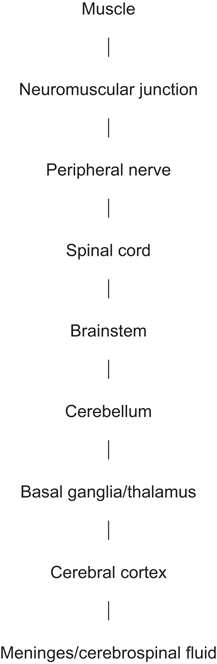
In defining the neuroanatomic site, it is helpful to establish the highest and lowest levels of the nervous system that can give rise to the patient’s signs and symptoms. Helpful keys in determining the most likely site include
1.
Find the earliest signs and symptoms of the illness, which usually denotes anatomically where the disease began.
2.
Determine the anatomic site where weakness and/or sensory changes likely are produced. Motor and sensory systems are multisynaptic long tract systems commonly involved in many diseases. For example, weakness from motor system dysfunction can occur at the motor cortex, brainstem, spinal cord, peripheral nerves, neuromuscular junctions, and/or muscle level—and each site has unique characteristics that help localize the problem.
3.
Identify accompanying non-neurologic signs and symptoms that may help localize site.
Although there are many neurologic signs and symptoms that point to a given neuroanatomic site, below are some of the more common clinical features—keeping in mind that there can be exceptions to these rules in certain diseases.
Muscle
Weakness without sensory loss
Proximal muscles weaker than distal muscles
Weakness often slowly progressive
Muscle atrophy
Neuromuscular Junction
Fatigue (especially in chewing and in proximal limb muscles)
Weakness without sensory loss
Ptosis with episodic diplopia
No muscle atrophy
Peripheral Nerve
Mixture of motor and sensory findings
Distribution of signs may be in a single nerve or multiple nerves
Distal limb signs more pronounced than proximal signs
Trunk uncommonly involved
Pain in feet or along a single nerve distribution
Sensory loss may be to pain and temperature, vibration and position sense, or multiple modalitiesRelated posts:

Stay updated, free articles. Join our Telegram channel

Full access? Get Clinical Tree


