28 Atypical Motor Neuron Disorders
There are a heterogeneous group of motor neuron disorders that are rare but nonetheless important to recognize, because they often can mimic the presentation of amyotrophic lateral sclerosis (ALS). These often are referred to as atypical motor neuron disorders. Although many of the atypical motor neuron disorders share some features with ALS, they often can be distinguished by their clinical and electrophysiologic characteristics (Boxes 28–1 and 28–2).
Box 28–1
Clinical Clues of An Atypical Motor Neuron Disorder
Non-myotomal pattern of weakness
Absence of significant muscle wasting in chronically weak limbs
Predominantly lower motor neuron signs
Presence of sensory symptoms and/or signs
Cerebellar, extrapyramidal, cognitive, and/or psychiatric dysfunction
Duration of illness longer than 5 years
Onset of illness before age 40 years
History of human immunodeficiency virus infection
Box 28–2
Electrodiagnostic Clues of An Atypical Motor Neuron Disorder
Conduction block on motor nerve conduction studies (not at entrapment sites)
Markedly slowed conduction velocities or prolonged distal latencies (not at entrapment sites)
Sensory nerve conduction abnormalities
Prominent complex repetitive discharges
Facial fasciculations/grouped repetitive motor unit discharges with activation
Acute or subacute neuropathic pattern on needle electromyogram
One of the most important atypical motor neuron disorders that can be confused with motor neuron disease is the immune-mediated motor neuropathy multifocal motor neuropathy with conduction block (MMNCB). Strictly speaking, this is a disorder of the motor nerve and as such is discussed in detail in Chapter 26. Patients present with progressive, asymmetric weakness and wasting that often affect the distal upper extremity muscles first. Weakness is in the distribution of named motor nerves, often with sparing of other nerves in the same myotome (clinical multifocal motor neuropathy). This pattern is not seen in ALS or its progressive muscular atrophy variant, in which the entire myotome is characteristically affected at the same time. Occasional patients have weakness without wasting, a finding usually associated with pure demyelination. The disease is slowly progressive, with a male predilection, generally presenting before the fifth decade. Definite upper motor neuron signs are absent, although retained or inappropriately brisk reflexes for the degree of weakness and wasting may be seen. Bulbar function and sensation are characteristically spared. Mild or transient sensory symptoms may be present. The characteristic finding on motor nerve conduction studies is that of conduction block, temporal dispersion, or both, along the motor nerves. Other signs of demyelination also may be seen, including slowed conduction velocities, absent or impersistent F responses, and prolonged distal motor latencies. Sensory conduction studies are typically normal.
Infectious Motor Neuron Disorders
West Nile Encephalitis
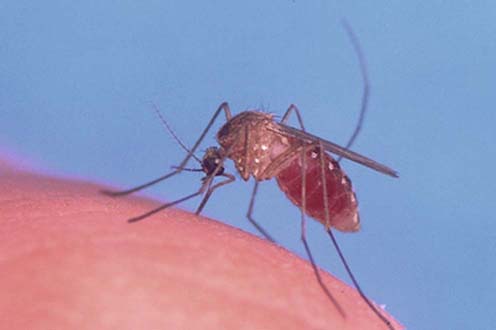
Over the past several years, there have been an increasing number of reports of a “polio-like” syndrome associated with West Nile encephalitis. The responsible virus, which is a member of the flavivirus family and is composed of a single strand of RNA, was first isolated in 1937 in northern Uganda. In nature, the virus is transmitted between birds by mosquitoes (Figure 28–1). Jays, blackbirds, finches, warblers, sparrows, and crows appear to be most important in maintaining the infection. Most infections in humans occur by way of a mosquito bite, although cases have been reported following transplanted organs and infected blood products. Because the disease is primarily spread to humans by mosquitoes, patients typically are affected in the summer and early fall.


