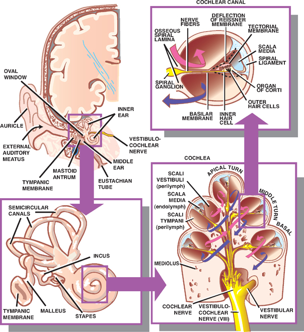
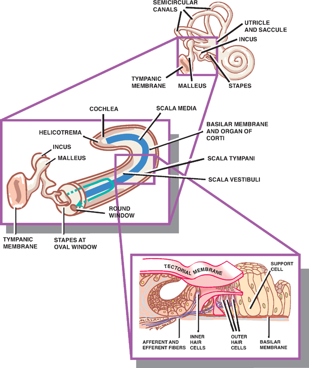
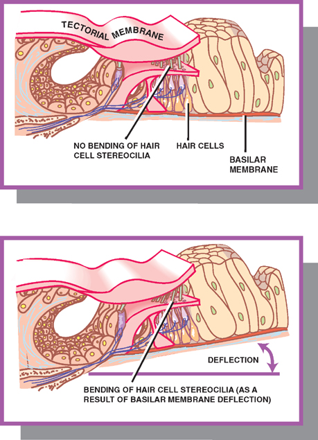
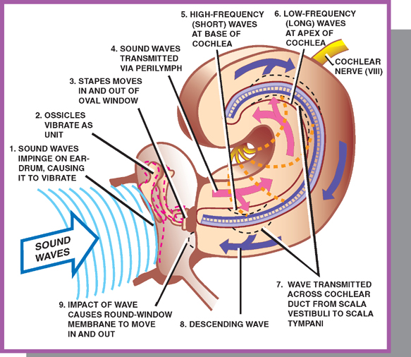
16 Auditory System The auditory system consists of the external ear, middle ear, cochlea of the inner ear, cochlear nerve, and central auditory pathways. See Fig. 16.1. The external ear comprises the auricle and the external auditory meatus. It is separated from the middle ear by the tympanic membrane. The middle ear comprises three main chambers: the tympanic cavity, the mastoid antrum, and the eustachian tube. The tympanic cavity contains three ossicles that transmit vibrations from the tympanic membrane to the auditory labyrinth. The malleus abuts the tympanic membrane, the stapes abuts the oval window, and the incus is set between the two. The mastoid antrum communicates with pneumatized spaces of the mastoid. The eustachian tube connects the tympanic cavity with the pharynx. Fig. 16.1 External, middle, and inner ear. See Figs. 16.1 and 16.2. The inner ear comprises two parts: a bony labyrinth formed by cavernous openings in the petrous portion of the temporal bone and a membranous labyrinth formed by a simple epithelial membrane. The membranous labyrinth, which is filled with a fluid called endolymph, lines the contours of the bony labyrinth from which it is separated by a fluid called perilymph. The membranous labyrinth consists of the cochlea, the utricle and saccule, and the semicircular canals. The cochlea, or auditory labyrinth, is responsible for hearing. The utricle and saccule and the semicircular canals make up the vestibular labyrinth. See Figs. 16.1 and 16.2. The cochlea is the auditory part of the membranous and bony labyrinth. It is shaped like the shell of a snail. It consists of (1) a bony core called the mediolus, which contains the cochlear part of the vestibulocochlear nerve, and (2) a cochlear canal, which winds two and one-half times around the mediolus like the threads of a screw. The cavity of the cochlear canal is divided into three compartments: scala vestibuli, scala tympani, and scala media. The scala media, which is situated in the middle portion of the cochlear canal, contains endolymph. The scala vestibuli and scala tympani, which are situated above and below the scala media, respectively, are filled with perilymph. The scala vestibuli and scala tympani communicate with each other at the apex of the cochlea called the helicotrema; the scala media ends in a blind sac. Reissner’s membrane separates the scala vestibuli from the scala media; the basilar membrane separates the scala tympani from the scala media. These membranes are attached to the bony wall of the cochlear canal by the spiral ligament. Fig. 16.2 Middle and inner ear. See Fig. 16.3. The organ of Corti is composed of sensory hair cells. It is situated on the scala media side of the basilar membrane. The hair cells of the organ of Corti contain stereo-cilia that project into an overlying gelatinous structure called the tectorial membrane. The sequence of steps involved in the transduction of fluid wave energy into electric action potentials is described as follows. Inward movement of the oval window by the stapes sets up a complicated fluid wave. The fluid wave travels along the scala vestibuli and scala tympani and across the scala media to cause upward displacement of the basilar membrane. As the basilar membrane is forced upward against a fxed tectorial membrane, shearing forces are exerted on the intervening stereocilia, causing excitation of the sensory hair cells. The point of maximum displacement along the basilar membrane is determined by the sound frequency transmitted: low-frequency sounds result in maximum displacement near the apex; high-frequency sounds result in maximum displacement near the base. Excitability of cochlear nerve fibers is also frequency dependent. Each fiber is most sensitive at a specific frequency. This spatial distribution of sound frequency within the cochlea is clinically manifest by cochlear lesions that result in threshold losses for certain parts of the auditory spectrum. Fig. 16.3 Organ of Corti. See Fig. 16.4. The mechanical events involved in the transduction of sound waves into chemoelectrical potentials occur as follows: Two features of the middle ear enhance the effciency of the energy transfer from air to fluid: (1) because the area of the oval window is substantially smaller than the area of the tympanic membrane, the vibratory force created by sound waves at the tympanic membrane is greatly magnified at the fluid interface of the oval window; (2) because fluid has a higher acoustic impedance than air, the direct transfer of sound waves to the fluid-filled cochlea would result in a substantial loss of energy. The transfer of sound energy to the fluid-filled cochlea via the tympanic membrane and ossicles ameliorates this potential loss of energy.
Functional Anatomy of the Auditory System
The External and Middle Ear

The Inner Ear
Cochlea

Organ of Corti

Conductive Hearing

Stay updated, free articles. Join our Telegram channel

Full access? Get Clinical Tree


