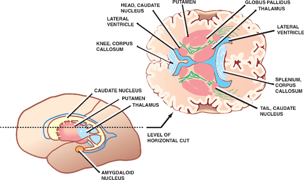
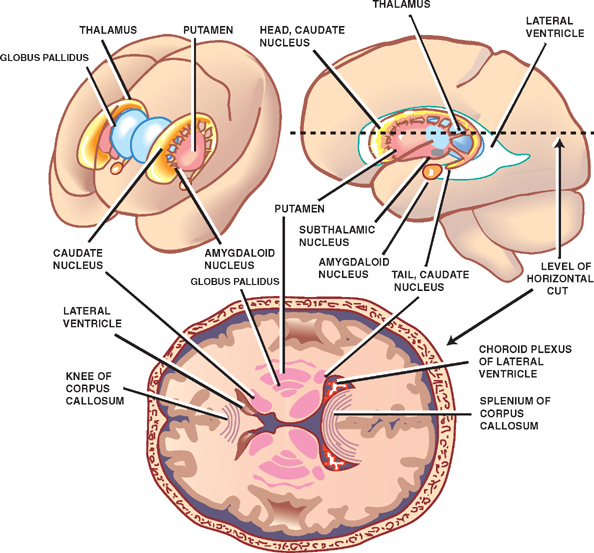
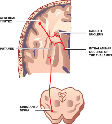
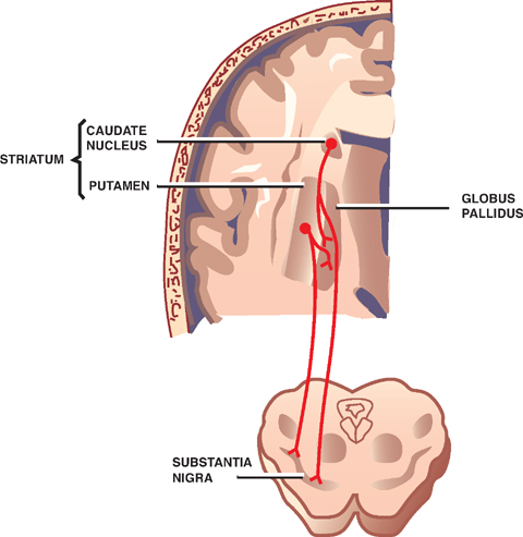
11 Basal Ganglia The basal ganglia are a collection of five subcortical nuclei located deep in the cerebral hemispheres. They play an important role in the control of posture and voluntary movement. Like the cerebellum, the basal ganglia are components of the motor system that serve to influence the major descending tracts (i.e., the corticospinal and corticobulbar tracts). Unlike the cerebellum, however, the basal ganglia have no direct connections with the spinal cord. Instead, they act as regulators of cortical function via their influence on thalamocortical projections. Thus, the major output of the basal ganglia is directed to the thalamus, which in turn projects to the frontal cortex; the major input is received from large cortical areas, forming a loop that may be summarized as follows: cortex-basal ganglia-thalamus-cortex. The main components of the basal ganglia involved in this loop are (1) the caudate nucleus, (2) the putamen, and (3) the globus pallidus. Two related subcortical nuclei traditionally included among the basal ganglia are (4) the subthalamic nucleus and (5) the substantia nigra. This chapter reviews the basic anatomy of the five main components of the basal ganglia, describes their main connections, and briefly reviews their neurochem-istry and physiology. In closing, the chapter briefly reviews the clinical manifestations of basal ganglia lesions, which have played an important role in our current understanding of basal ganglia function. See Figs. 11.1 and 11.2. As noted, the basal ganglia comprise five subcortical nuclei: (1) the caudate nucleus, (2) the putamen, (3) the globus pallidus, (4) the subthalamic nucleus, and (5) the substantia nigra. The caudate nucleus and the putamen have many functional and developmental similarities and are usually considered together as a single structure, the striatum. The striatum is composed of the caudate nucleus and the putamen. Both nuclei receive projections from the telen-cephalon, and together they constitute the input component of the basal ganglia. The caudate nucleus is C-shaped and is closely related to the lateral ventricle. It lies lateral to the thalamus and medial to the fibers of the internal capsule. It is composed of three parts, which are related to the three main areas of the lateral ventricle, as follows: In contrast to the striatum, the globus pallidus derives from the diencephalon. It is divided into external and internal parts. The latter constitutes the output component of the basal ganglia. The globus pallidus is the smallest nucleus of the basal ganglia. Laterally, it is bordered by the putamen. Medially, it is bordered by the internal capsule. Together, the putamen and globus pallidus form a lens-shaped structure, which is sometimes called the lentiform nucleus—a purely structural designation not employed here. The subthalamic nucleus forms a side loop with the globus pallidus. It is a lens-shaped nucleus located on the medial side of the internal capsule at the border between the diencephalon and the mesencephalon. Caudally, it is continuous with the substantia nigra. The substantia nigra is located in the mesencephalon. It is divided into two regions: a dorsal pars compacta and a ventral pars reticulata. These two regions have distinct histological and functional characteristics. The pars compacta is composed of neurons that contain the pigment melanin, giving the substantia nigra its dark color which is reflected in its name. These neurons project rostrally and form a side loop with the striatum. The pars reticulata is directly continuous with the globus pallidus with which it shares both histological and functional characteristics. Fig. 11.1 Basal ganglia. Fig. 11.2 Striatum and globus pallidus. All input into the basal ganglia terminates in the stria-tum; all output projects from the globus pallidus and the pars reticulata of the substantia nigra. See Fig. 11.3. There are three major sources of input to the striatum: Fig. 11.3 Afferent connections of the striatum. See Fig. 11.4. There are two major destinations of striatal efferents:
Components of the Basal Ganglia
Striatum
Globus Pallidus
Subthalamic Nucleus
Substantia Nigra
Connections of the Basal Ganglia
Striatum
Afferents
Efferents
Related posts:

Stay updated, free articles. Join our Telegram channel

Full access? Get Clinical Tree


