Fig. 27.1
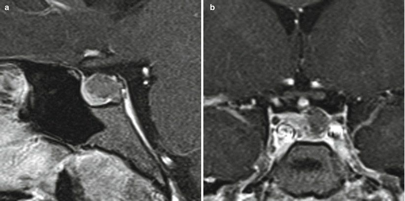
Colloid cyst. (a) Sagittal T1-weighted post-gadolinium image. (b) Coronal T1-weighted post-gadolinium image. A lobular cyst with a thin rim of enhancement is centered in the sella, with suprasellar extension. The sellar floor is remodeled. The optic chiasm appears to be elevated by the cyst

Fig. 27.2
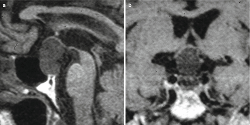
Colloid cyst. (a) Sagittal T1-weighted post-gadolinium image. (b) Coronal T1-weighted post-gadolinium image. A slightly hyperintense cyst is centered in the left sella, displacing the normal pituitary tissue to the left. The sellar floor is expanded

Fig. 27.3
Colloid cyst. (a) Sagittal T1-weighted nonenhanced image. (b) Coronal T1-weighted nonenhanced image. A lobular cyst with a thin rim of enhancement is centered in the sella, with suprasellar extension. The sellar floor is remodeled. The optic chiasm appears to be elevated by the cyst
27.3 Histopathology
Colloid cysts are typically filled with a whitish or yellowish, relatively thick, colloid (mucopolysaccharide) substance. Areas of old hemorrhage and cholesterol accumulation may give the contents of the cyst a denser consistency.
There may be a dense, calcified component within the cyst.
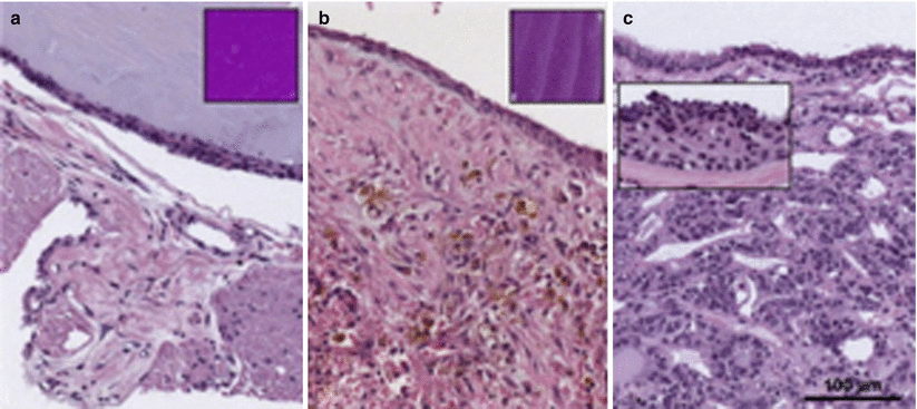
Histologically, colloid cysts are lined by a single layer of cuboidal or columnar epithelium with interspersed ciliated and goblet cells (Fig. 27.4).
Colloid cysts are often indistinguishable from RCCs; many consider them the same entity [6].


