Fig. 58.1
Neuromyelitis optica (NMO). (a) Coronal fat-saturated T1-weighted gadolinium-enhanced MR image. (b) Coronal inversion recovery image. (c) Sagittal T1-weighted gadolinium-enhanced image. The left optic nerve demonstrates edema (b) and enhancement (a) consistent with neuritis. There is also focal enhancement of the lower thoracic cord (c) (arrow)
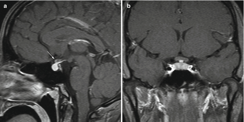
Fig. 58.2
NMO. (a) Sagittal T1-weighted gadolinium-enhanced MR image. (b) Coronal T1-weighted gadolinium-enhanced image. This young woman with vision loss and hypopituitarism was found to have NMO. There is slight enhancement of the optic chiasm (arrow) and infundibulum
MRI in some patients with NMO is normal.
Optical coherence tomography (OCT) may be used to measure the thickness of the retinal nerve fiber layer in MS and NMO.
58.3 Histopathology
White matter plaques with demyelination and extensive perivenous infiltration of lymphocytes, macrophages, and plasma cells are commonly seen.
Axonal tracts are typically preserved.
Astrogliosis is often seen.
In severe cases, necrotizing inflammation of the white matter can be seen.
IgG and IgM antibodies are often positive in affected tissue.
58.4 Clinical Management
Although not all patients with NMO are seroconverted, serum evidence of aquaporin-4 antibodies confirm the diagnosis (NMO-IgG antibody). This test has a sensitivity of 75 % in diagnosing NMO [18].
Diagnostic criteria for NMO currently require optic neuritis, acute myelitis, and two of the following three characteristics [19]:
MRI that is nondiagnostic for MS
A longitudinal spinal cord lesion extending over three vertebral segments
NMO-IgG seropositive status
Treatment for acute NMO includes high-dose corticosteroids and plasmapheresis.
Long-term treatment may involve drugs such as azathioprine, mofetil mycophenolate, and rituximab [20].
A frequent relapsing course is observed in over 80 % of patients with NMO [19].
References
1.
2.
3.
Huitinga I, Erkut ZA, van Beurden D, Swaab DF. Impaired hypothalamus-pituitary-adrenal axis activity and more severe multiple sclerosis with hypothalamic lesions. Ann Neurol. 2004;55:37–45.CrossRefPubMed
Related posts:


Stay updated, free articles. Join our Telegram channel

Full access? Get Clinical Tree


