Fig. 72.1
Traumatic injury to anterior skull base fracture. (a) Sagittal CT image reconstructed in bone algorithm. (b) Coronal CT image with intravenous contrast enhancement. Comminuted fractures of the planum sphenoidale are seen on both images (arrows), with complete opacification of the sphenoid sinuses and pneumocephalus (a, upper arrow)
On MRI, enlargement of the pituitary gland may be seen. Specific changes within the pituitary gland, such as hemorrhage or infarction, are seen in 30 % of cases following TBI [17].
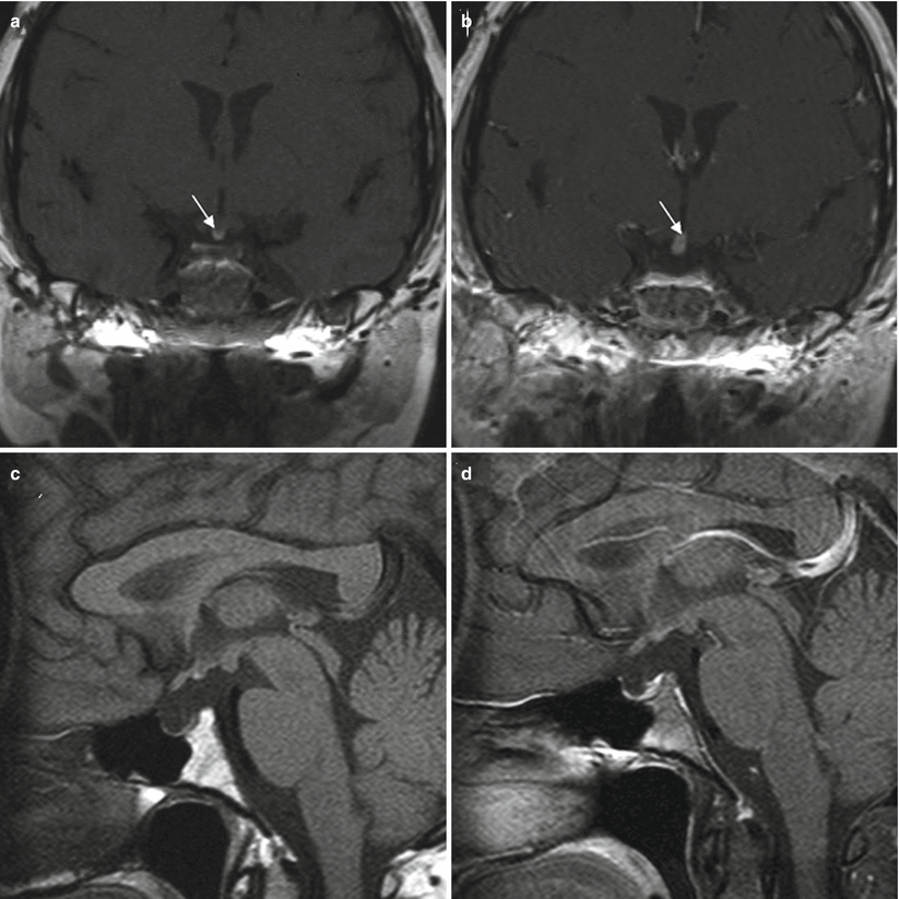
MRI may show pituitary stalk disruption with loss of the normal posterior pituitary bright spot; this loss has been reported to correlate clinically with the development of diabetes insipidus (Fig. 72.2) [7, 18, 19].


Fig. 72.2
Pituitary stalk transection. (a, b) Coronal pituitary MR images before and after contrast administration in a patient with pituitary stalk transection. Residual pituitary (arrows) is seen at the floor of the third ventricle. The figure also illustrates the small anterior pituitary gland and absent infundibulum. (c, d) Sagittal pituitary MR images before and after contrast administration (Adapted with permission from Ioachimescu et al. [19])
In up to 80 % of long-term survivors of TBI with clinical hypopituitarism, MRI may show an empty sella, perfusion deficits, and loss of the posterior pituitary bright spot [20].
In patients with posttraumatic anosmia, MR imaging is often negative, or it may show diffuse white matter changes consistent with prior TBI. The orbitofrontal cortex is most likely to show evidence of injury in patients with anosmia.
SPECT imaging has been used to measure perfusion, which may be altered in the frontal lobes in patients with posttraumatic anosmia [21].
72.3 Histopathology
Microscopic histological analysis of the pituitary gland following severe TBI shows a pattern of infarction and widespread necrosis of the anterior pituitary gland [22].
72.4 Clinical and Surgical Management
Standard neurocritical management in patients with TBI includes monitoring and treatment of intracranial pressure, supportive care, seizure control, and evacuation of any compressive lesions.
Management of traumatic CSF leaks should be performed in a timely fashion (in stable TBI patients) to prevent pneumocephalus or meningitis.
Screening for hypopituitarism is recommended for patients with severe TBI, especially children, older adults, and patients with evidence of basilar skull fractures. Some patients will require additional stress doses of hormones that they are unable to produce in a posttraumatic state, and growth hormone deficiency has more profound effects on children than on adults.
In some patients with visual loss following TBI, decompression of the optic nerve or chiasm is recommended. This can be done via a craniotomy or endoscopic endonasal approach. Approximately one third to one half of patients will have improvement in vision following surgical decompression of the optic nerve. Steroids do not appear to provide an additional clinical benefit [8, 25, 26].
In patients with posttraumatic anosmia, the key aspects of clinical management involve recognizing the anosmia and treating any associated conditions, including CSF leaks, infections, and neuropsychiatric dysfunction.
Patients with craniofacial trauma and anosmia are more likely to have psychosocial and vocational dysfunction for years following an injury; these patients may need multidisciplinary treatment involving therapists, psychiatrists, and others.
Recovery of olfactory function has been reported in 10 % of patients, occurring between 8 weeks and 2 years after the injury [27].
References
1.
2.
Schneider M, Schneider HJ, Yassouridis A, Saller B, von Rosen F, Stalla GK. Predictors of anterior pituitary insufficiency after traumatic brain injury. Clin Endocrinol (Oxf). 2008;68:206–12.
3.
Tanriverdi F, De Bellis A, Bizzarro A, Sinisi AA, Bellastella G, Pane E, et al. Antipituitary antibodies after traumatic brain injury: is head trauma-induced pituitary dysfunction associated with autoimmunity? Eur J Endocrinol. 2008;159:7–13.CrossRefPubMed
Stay updated, free articles. Join our Telegram channel

Full access? Get Clinical Tree


