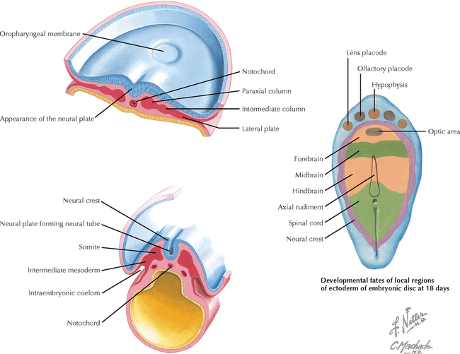
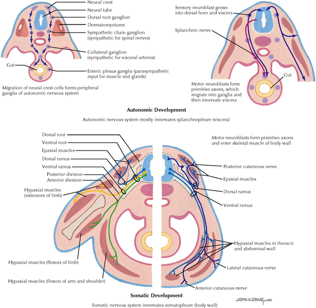
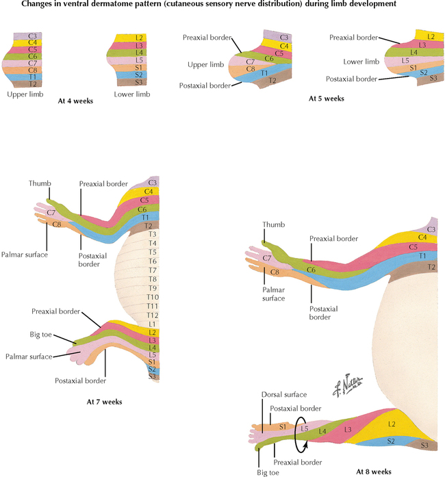
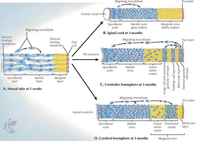
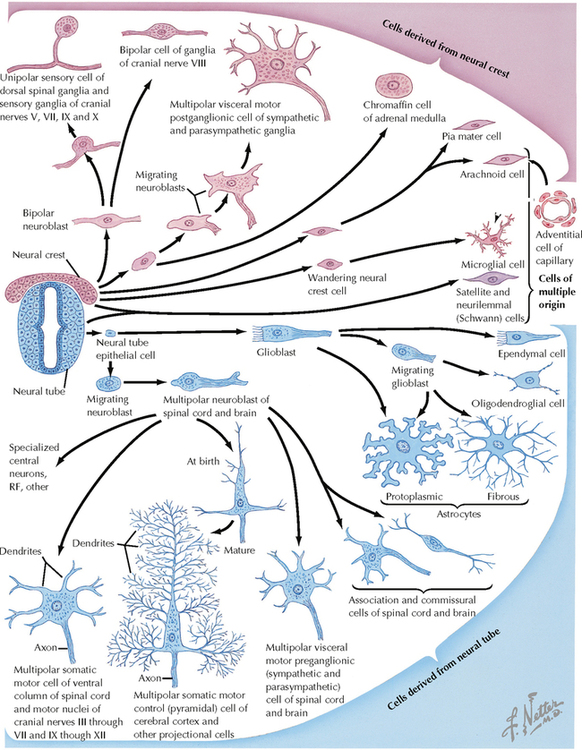
8 DEVELOPMENTAL NEUROSCIENCE 8.1. Formation of the Neural Plate, Neural Tube, and Neural Crest 8.2. Neurulation 8.3. Neural Tube Development and Neural Crest Formation 8.4. Development of Peripheral Axons 8.5. Somatic Versus Splanchnic Nerve Development 8.6. Limb Rotation and Dermatomes 8.7. Neural Proliferation and Differentiation: Walls of the Neural Tube 8.8. Neural Tube and Neural Crest Derivatives 8.9. Early Brain Development: The 28-Day-Old Embryo 8.10. Early Brain Development: The 36-Day-Old Embryo 8.11. Early Brain Development: The 49-Day-Old Embryo and the 3-Month-Old Embryo 8.12. Forebrain Development: 7 Weeks through 3 Months 8.13. The 6-Month and 9-Month Central Nervous Systems 8.14. Comparison of 5½ Week and Adult Central Nervous System Regions 8.15. Alar and Basal Plate Derivatives in the Brain Stem 8.16. Adult Derivatives of the Forebrain, Midbrain, and Hindbrain 8.17. Cranial Nerve Primordia 8.18. Cranial Nerve Neuron Components 8.19. Development of Motor and Preganglionic Autonomic Nuclei in the Brain Stem and Spinal Cord 8.20. Development of the Eye and Orbit 8.21. Development of the Ear 8.22. Development of the Pituitary Gland 8.23. Development of the Ventricles 8.24. Development of the Fourth Ventricule 8.25. Neural Tube Defects 8.26. Defects of the Brain and Skull 8.1 FORMATION OF THE NEURAL PLATE, NEURAL TUBE, AND NEURAL CREST The neural plate, neural tube, and neural crest form at the 18-day stage of embryonic development. The underlying notochord induces the neural plate, and a midline neural groove forms. The elevated lateral margins become the neural folds, tissue destined to become the neural crest with future contributions to many components of the peripheral nervous system (PNS). At this very early stage of embryonic development, these neural precursors are vulnerable to toxic and other forms of insult. 8.2 NEURULATION In the 21- or 22-day-old-embryo, the neural plate, with its midline neorural groove, thickens and begins to fold and elevate along either side, allowing the two lateral edges to fuse at the dorsal midline to form the completed neural tube. The central canal, the site of the future development of the ventricular system, is in the center of the neural tube. This process of neurulation continues both caudally and rostrally. Disruption can occur because of failure of full neural tube formation caudally (spina bifida) or rostrally (anencephaly). CLINICAL POINT As the neural plate forms into a neural tube, the process of neurulation results in fused neural folds, starting centrally and moving both caudally and rostrally. Failure of the neural tube to close results in dysraphic defects, with altered development of associated muscles, bone, skin, and meninges. If the anterior neuropore fails to form, anencephaly results, with failure of the brain to develop, accompanied by facial defects. This condition is lethal. Failure of the posterior (caudal) neuropore to close results in spina bifida, with failure of the vertebral arches to fuse. A saccular protrusion from the lumbar region may contain meninges (meningocele) or meninges and spinal cord (meningomyelocele). Meningomyelocele is often accompanied by paraparesis, bowel and bladder dysfunction, sensory disruption at the level of the lesion, and accompanying hydrocephalus or Arnold-Chiari malformation, requiring a ventriculo-peritoneal or ventriculo-jugular shunt. 8.3 NEURAL TUBE DEVELOPMENT AND NEURAL CREST FORMATION The dorsal and ventral halves of the neural tube are separated by the sulcus limitans, an external protrusion from the central canal that demarcates the alar plate above from the basal plate below. This important landmark persists at some sites in the adult ventricular system. The alar plate is the source of generation of many neurons with sensory function. The basal plate is the source of generation of many neurons with motor or autonomic function in the spinal cord and the brain stem. The neural crest cells at the edge of the neural folds unite and become a dorsal crest, the neural crest above the neural tube. The neural tube and neural crest separate from the originating ectoderm. CLINICAL POINT The neural crest gives rise to a wide variety of neural elements of the PNS, including primary sensory neurons, postganglionic autonomic neurons, Schwann cells, adrenal medullary chromaffin cells, pia and arachnoid cells, melanocytes, and some mesenchyme of the head. A failure of the neural crest to develop and migrate properly is seen in Hirschsprung’s disease (congenital megacolon), in which sensory signals from the colon are absent, and in familial dysautonomia, in which autonomic symptoms (cardiovascular dysfunction, gastrointestinal dysfunction) and sensory deficits (especially pain and temperature sensation) are present. 8.4 DEVELOPMENT OF PERIPHERAL AXONS Peripheral axon development is a complex process of central and peripheral neurite extension, trophic and chemotactic factors, and axonal guidance and maintenance by innervated target tissues. Dorsal root ganglion cells are bipolar; a peripheral axonal process is associated with simple or complex sensory receptor cells, and a central axonal process extends into the central nervous system (CNS) to form connections with secondary sensory neurons. The lower motor neurons send motor axons to the developing skeletal muscles through the ventral roots or motor cranial nerves, forming neuromuscular junctions as sites of synaptic connectivity. Motor neurons that fail to establish such contact with skeletal muscles die. Central preganglionic axons exit in the ventral roots and terminate on sympathetic ganglion cells in the sympathetic chain or collateral ganglia or on parasympathetic intramural ganglia near the organs innervated. Postganglionic axons form connections with target tissues, including smooth muscle, cardiac muscle, secretory glands, some metabolic cells (hepatocytes, fat cells), and cells of the immune system in parenchymal zones of many lymphoid organs. Sensory, motor, and autonomic symptoms can occur in peripheral neuropathies based on disruption of these connections. 8.5 SOMATIC VERSUS SPLANCHNIC NERVE DEVELOPMENT Somatopleure and splanchnopleure constitute the embryonic basis for the subdivision of the PNS into spinal (somatic) nerves and splanchnic (autonomic) nerves. The somatopleure develops from ectoderm and the somatic portion of lateral plate mesoderm. Somite hypoblasts migrate into somatopleure to form the lateral and ventral aspects of the body wall, including the limbs. Splanchnopleure, derived from endoderm and lateral plate mesoderm, give rise to visceral organs. The ventral rami migrate into somatopleure, and splanchnic nerves grow into splanchnopleure. Thoracic and lumbar splanchnic nerves have sympathetic and visceral sensory axonal components. Pelvic splanchnic nerves (S2–S4) have parasympathetic and visceral sensory axonal components. 8.6 LIMB ROTATION AND DERMATOMES Rotation of the lower limb results in a reversal of the preaxial and postaxial borders, producing a spiral arrangement of dermatomes. Spinal nerve segments on the anterior surface of the lower extremity extend medially and inferiorly; the great toe (hallux) is supplied by nerves from a more rostral dermatome (L4) than the little toe (S1). The lower extremity is an extension of the trunk, and the most caudal dermatomes (sacral and coccygeal) supply the perineum, not the foot. Cervical dermatomes maintain a relatively orderly distribution to the upper extremity with minimal rotation. 8.7 NEURAL PROLIFERATION AND DIFFERENTIATION: WALLS OF THE NEURAL TUBE Early in development (5 weeks), neuroblasts in the ependymal layer lining the central canal move back and forth from the ependymal surface to the pial surface, replicating as they go. Neural migration follows distinctive patterns in different regions of the neural tube. In the spinal cord, neurons migrate into the inner mantle zone, leaving the outer marginal zone as a site for axonal pathways. In the cerebellar cortex, some neurons migrate to an outer location on the outer pial surface as an external granular layer, from which granular cells then migrate inward to synapse with other neurons present in deeper layers of the cerebellar cortex. In the cerebral cortex, neurons migrate to the outer zone, where the gray matter (neuronal cell bodies) remains on the surface, external to the white matter (nerve fibers). These developmental patterns reflect the anatomical organization of the mature structures, their blood supply, and their vulnerability to injury by tumors, vascular insults, trauma, and other disorders. 8.8 NEURAL TUBE AND NEURAL CREST DERIVATIVES Only gold members can continue reading. Log In or Register to continue Share this:Click to share on Twitter (Opens in new window)Click to share on Facebook (Opens in new window) Related Related posts: VENTRICLES AND THE CEREBROSPINAL FLUID SPINAL CORD MOTOR SYSTEMS AUTONOMIC-HYPOTHALAMIC-LIMBIC SYSTEMS TELENCEPHALON PERIPHERAL NERVOUS SYSTEM Stay updated, free articles. Join our Telegram channel Join Tags: Netters Atlas of Neuroscience with STUDENT CONSULT Online Access Jun 4, 2016 | Posted by admin in NEUROLOGY | Comments Off on DEVELOPMENTAL NEUROSCIENCE Full access? Get Clinical Tree
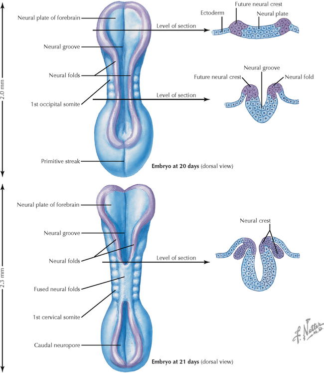
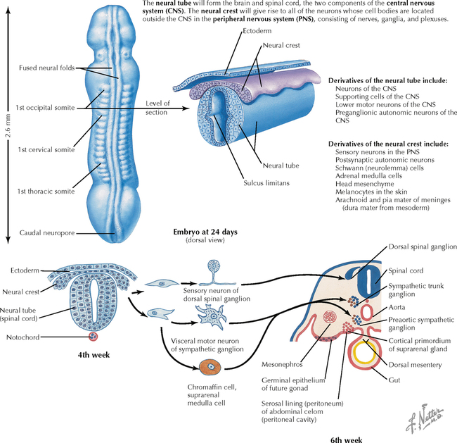
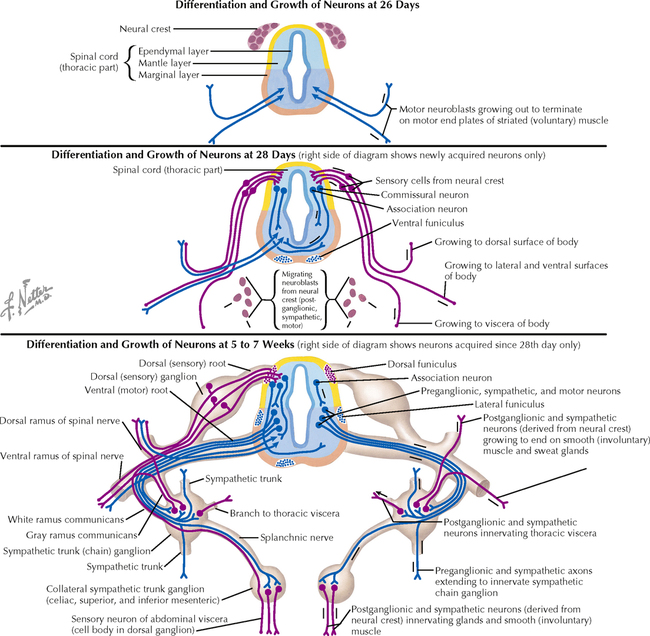
8 DEVELOPMENTAL NEUROSCIENCE 8.1. Formation of the Neural Plate, Neural Tube, and Neural Crest 8.2. Neurulation 8.3. Neural Tube Development and Neural Crest Formation 8.4. Development of Peripheral Axons 8.5. Somatic Versus Splanchnic Nerve Development 8.6. Limb Rotation and Dermatomes 8.7. Neural Proliferation and Differentiation: Walls of the Neural Tube 8.8. Neural Tube and Neural Crest Derivatives 8.9. Early Brain Development: The 28-Day-Old Embryo 8.10. Early Brain Development: The 36-Day-Old Embryo 8.11. Early Brain Development: The 49-Day-Old Embryo and the 3-Month-Old Embryo 8.12. Forebrain Development: 7 Weeks through 3 Months 8.13. The 6-Month and 9-Month Central Nervous Systems 8.14. Comparison of 5½ Week and Adult Central Nervous System Regions 8.15. Alar and Basal Plate Derivatives in the Brain Stem 8.16. Adult Derivatives of the Forebrain, Midbrain, and Hindbrain 8.17. Cranial Nerve Primordia 8.18. Cranial Nerve Neuron Components 8.19. Development of Motor and Preganglionic Autonomic Nuclei in the Brain Stem and Spinal Cord 8.20. Development of the Eye and Orbit 8.21. Development of the Ear 8.22. Development of the Pituitary Gland 8.23. Development of the Ventricles 8.24. Development of the Fourth Ventricule 8.25. Neural Tube Defects 8.26. Defects of the Brain and Skull 8.1 FORMATION OF THE NEURAL PLATE, NEURAL TUBE, AND NEURAL CREST The neural plate, neural tube, and neural crest form at the 18-day stage of embryonic development. The underlying notochord induces the neural plate, and a midline neural groove forms. The elevated lateral margins become the neural folds, tissue destined to become the neural crest with future contributions to many components of the peripheral nervous system (PNS). At this very early stage of embryonic development, these neural precursors are vulnerable to toxic and other forms of insult. 8.2 NEURULATION In the 21- or 22-day-old-embryo, the neural plate, with its midline neorural groove, thickens and begins to fold and elevate along either side, allowing the two lateral edges to fuse at the dorsal midline to form the completed neural tube. The central canal, the site of the future development of the ventricular system, is in the center of the neural tube. This process of neurulation continues both caudally and rostrally. Disruption can occur because of failure of full neural tube formation caudally (spina bifida) or rostrally (anencephaly). CLINICAL POINT As the neural plate forms into a neural tube, the process of neurulation results in fused neural folds, starting centrally and moving both caudally and rostrally. Failure of the neural tube to close results in dysraphic defects, with altered development of associated muscles, bone, skin, and meninges. If the anterior neuropore fails to form, anencephaly results, with failure of the brain to develop, accompanied by facial defects. This condition is lethal. Failure of the posterior (caudal) neuropore to close results in spina bifida, with failure of the vertebral arches to fuse. A saccular protrusion from the lumbar region may contain meninges (meningocele) or meninges and spinal cord (meningomyelocele). Meningomyelocele is often accompanied by paraparesis, bowel and bladder dysfunction, sensory disruption at the level of the lesion, and accompanying hydrocephalus or Arnold-Chiari malformation, requiring a ventriculo-peritoneal or ventriculo-jugular shunt. 8.3 NEURAL TUBE DEVELOPMENT AND NEURAL CREST FORMATION The dorsal and ventral halves of the neural tube are separated by the sulcus limitans, an external protrusion from the central canal that demarcates the alar plate above from the basal plate below. This important landmark persists at some sites in the adult ventricular system. The alar plate is the source of generation of many neurons with sensory function. The basal plate is the source of generation of many neurons with motor or autonomic function in the spinal cord and the brain stem. The neural crest cells at the edge of the neural folds unite and become a dorsal crest, the neural crest above the neural tube. The neural tube and neural crest separate from the originating ectoderm. CLINICAL POINT The neural crest gives rise to a wide variety of neural elements of the PNS, including primary sensory neurons, postganglionic autonomic neurons, Schwann cells, adrenal medullary chromaffin cells, pia and arachnoid cells, melanocytes, and some mesenchyme of the head. A failure of the neural crest to develop and migrate properly is seen in Hirschsprung’s disease (congenital megacolon), in which sensory signals from the colon are absent, and in familial dysautonomia, in which autonomic symptoms (cardiovascular dysfunction, gastrointestinal dysfunction) and sensory deficits (especially pain and temperature sensation) are present. 8.4 DEVELOPMENT OF PERIPHERAL AXONS Peripheral axon development is a complex process of central and peripheral neurite extension, trophic and chemotactic factors, and axonal guidance and maintenance by innervated target tissues. Dorsal root ganglion cells are bipolar; a peripheral axonal process is associated with simple or complex sensory receptor cells, and a central axonal process extends into the central nervous system (CNS) to form connections with secondary sensory neurons. The lower motor neurons send motor axons to the developing skeletal muscles through the ventral roots or motor cranial nerves, forming neuromuscular junctions as sites of synaptic connectivity. Motor neurons that fail to establish such contact with skeletal muscles die. Central preganglionic axons exit in the ventral roots and terminate on sympathetic ganglion cells in the sympathetic chain or collateral ganglia or on parasympathetic intramural ganglia near the organs innervated. Postganglionic axons form connections with target tissues, including smooth muscle, cardiac muscle, secretory glands, some metabolic cells (hepatocytes, fat cells), and cells of the immune system in parenchymal zones of many lymphoid organs. Sensory, motor, and autonomic symptoms can occur in peripheral neuropathies based on disruption of these connections. 8.5 SOMATIC VERSUS SPLANCHNIC NERVE DEVELOPMENT Somatopleure and splanchnopleure constitute the embryonic basis for the subdivision of the PNS into spinal (somatic) nerves and splanchnic (autonomic) nerves. The somatopleure develops from ectoderm and the somatic portion of lateral plate mesoderm. Somite hypoblasts migrate into somatopleure to form the lateral and ventral aspects of the body wall, including the limbs. Splanchnopleure, derived from endoderm and lateral plate mesoderm, give rise to visceral organs. The ventral rami migrate into somatopleure, and splanchnic nerves grow into splanchnopleure. Thoracic and lumbar splanchnic nerves have sympathetic and visceral sensory axonal components. Pelvic splanchnic nerves (S2–S4) have parasympathetic and visceral sensory axonal components. 8.6 LIMB ROTATION AND DERMATOMES Rotation of the lower limb results in a reversal of the preaxial and postaxial borders, producing a spiral arrangement of dermatomes. Spinal nerve segments on the anterior surface of the lower extremity extend medially and inferiorly; the great toe (hallux) is supplied by nerves from a more rostral dermatome (L4) than the little toe (S1). The lower extremity is an extension of the trunk, and the most caudal dermatomes (sacral and coccygeal) supply the perineum, not the foot. Cervical dermatomes maintain a relatively orderly distribution to the upper extremity with minimal rotation. 8.7 NEURAL PROLIFERATION AND DIFFERENTIATION: WALLS OF THE NEURAL TUBE Early in development (5 weeks), neuroblasts in the ependymal layer lining the central canal move back and forth from the ependymal surface to the pial surface, replicating as they go. Neural migration follows distinctive patterns in different regions of the neural tube. In the spinal cord, neurons migrate into the inner mantle zone, leaving the outer marginal zone as a site for axonal pathways. In the cerebellar cortex, some neurons migrate to an outer location on the outer pial surface as an external granular layer, from which granular cells then migrate inward to synapse with other neurons present in deeper layers of the cerebellar cortex. In the cerebral cortex, neurons migrate to the outer zone, where the gray matter (neuronal cell bodies) remains on the surface, external to the white matter (nerve fibers). These developmental patterns reflect the anatomical organization of the mature structures, their blood supply, and their vulnerability to injury by tumors, vascular insults, trauma, and other disorders. 8.8 NEURAL TUBE AND NEURAL CREST DERIVATIVES Only gold members can continue reading. Log In or Register to continue Share this:Click to share on Twitter (Opens in new window)Click to share on Facebook (Opens in new window) Related Related posts: VENTRICLES AND THE CEREBROSPINAL FLUID SPINAL CORD MOTOR SYSTEMS AUTONOMIC-HYPOTHALAMIC-LIMBIC SYSTEMS TELENCEPHALON PERIPHERAL NERVOUS SYSTEM Stay updated, free articles. Join our Telegram channel Join Tags: Netters Atlas of Neuroscience with STUDENT CONSULT Online Access Jun 4, 2016 | Posted by admin in NEUROLOGY | Comments Off on DEVELOPMENTAL NEUROSCIENCE Full access? Get Clinical Tree