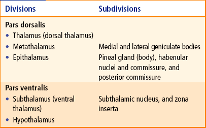
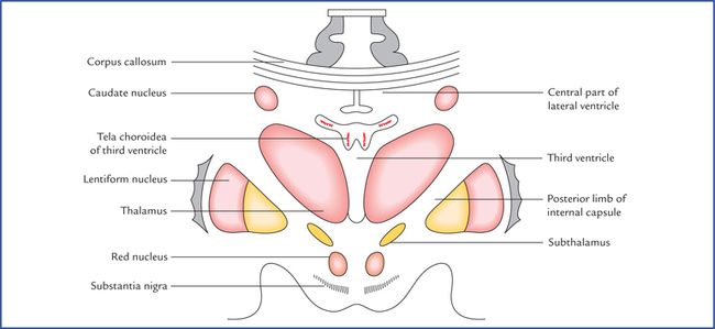
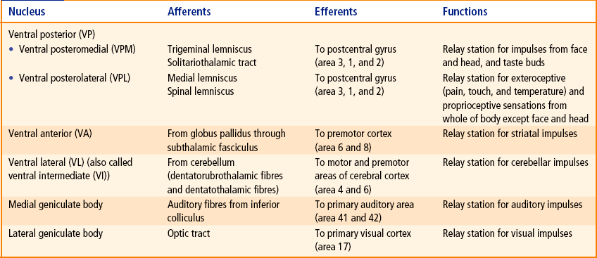
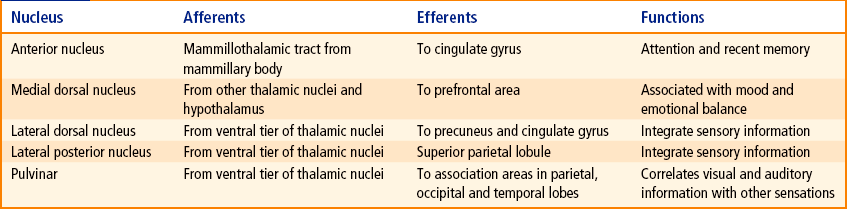
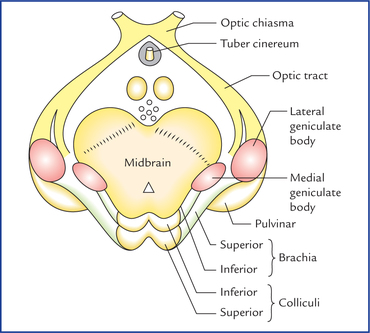
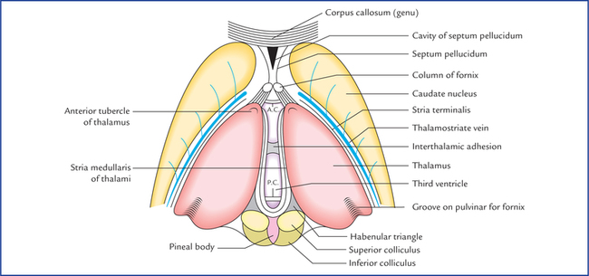
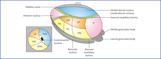
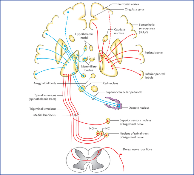
11 The diencephalon is the part of brain between the cerebrum and the brainstem. The cavity within it is termed third ventricle. The diencephalon comprises two major subdivisions: pars dorsalis and pars ventralis. These subdivisions are seen on midsagittal view of the brain, and are separated from each other by a shallow groove, the hypothalamic sulcus which extends from interventricular foramen to the rostral end of the cerebral aqueduct of midbrain (Fig. 11.10). • Pars dorsalis lies above (dorsal) to the hypothalamic sulcus and consists of: (a) thalamus, (b) metathalamus which includes the medial and lateral geniculate bodies, and (c) epithalamus which consists of pineal body (gland), habenular nuclei and commissure, posterior commissure and the stria medullaris thalami. • Pars ventralis lies below (ventral) to the hypothalamic sulcus and consists of: (a) subthalamus, and (b) hypothalamus. The main divisions and subdivisions of diencephalon are listed in Table 11.1. Anatomically the thalamus is a large ovoid mass of grey matter laying above the midbrain, from which it is separated by a small amount of neural tissue, the subthalamus. There are two thalami situated one on each side of a slitlike cavity, the third ventricle (Fig. 11.1). Fig. 11.1 The thalami and third ventricle as seen from above, after removal of overlying tela choroidea. (AC = anterior commissure, PC = posterior commissure.) Each thalamus is 3.5 cm in length and 1.5 cm in breadth. The long axes of the thalami are set obliquely running backwards and laterally. The pointed anterior ends are nearer to the median plane whereas the wider posterior ends are separated from each other by pineal body, superior colliculi and habenular triangles. The thalami are usually attached across the median plane by a narrow interthalamic connexus of grey matter (also called interthalamic adhesion). Each thalamus forms most of the lateral wall of the third ventricle and floor of the central part of the lateral ventricle. Each thalamus has two ends and four surfaces. • Superior surface. Its lateral part forms the floor of the central part of the lateral ventricle and its medial part is covered by the tela choroidea of the third ventricle. • Inferior surface. Its anterior part is fused with the sub-thalamus while its posterior part is free, forming the inferior aspect of the pulvinar. • Medial surface. It forms the greater part of the lateral wall of the third ventricle. • Lateral surface. It forms the medial boundary of the posterior limb of internal capsule. The thalamus consists mainly of grey matter and only a small amount of white matter. Fig. 11.3 Horizontal section of the thalamus (schematic) to show the location of various thalamic nuclei. The diagram in the inset is the coronal section of thalamus passing in front of pulvinar showing ventral posteromedial (VPM), ventral posterolateral (VPL) nuclei, and centromedian nucleus. (P = pulvinar, LD = lateral dorsal nucleus, LP = lateral posterior nucleus, VA = ventral anterior nucleus, VL = ventral lateral nucleus, VPL = ventral posterolateral nucleus, MN = Medio- dorsal nucleus.) A vertical Y-shaped sheet of white matter within the thalamus is called internal medullary lamina. The nuclei in this part are collectively referred to as anterior nucleus. Nuclei in medial part consist of a large medial dorsal nucleus and a small medial ventral nucleus. In addition to the above mentioned nuclei, the thalamus consists of following other nuclei: • Intralaminar nuclei. They are several in numbers and are embedded in the internal medullary lamina. The largest and most important of these is termed centrome-dian nucleus. • Midline (paraventricular) nuclei. They consist of scattered cells that lie between the medial part of the thala-mus and the ependyma of the third ventricle. • Reticular nucleus. It is a thin curved sheet of grey matter on the lateral aspect of the thalamus from which it is separated by the external medullary lamina. • Medial and lateral geniculate bodies. These are located posteroventral to the pulvinar. Conventionally these nuclei are described under metathalamus, but nowadays they are considered as thalamic nuclei. The thalamic nuclei are summarized in Table 11.2. Table 11.2 Nuclei in different parts of the thalamus These nuclei receive input from certain ascending tracts and project it to the specific (primary) cortical areas. Nuclei of this group comprise ventral tier nuclei and medial and lateral geniculate bodies. Their connections are enumerated in Table 11.3. Fig. 11.4 Main connections of the thalamus. The afferent fibres are shown on the left side and the efferent, fibres on the right side. (NG = nucleus gracilis, NC = nucleus cuneatus.) Fig. 11.5 Scheme to show connections of ventral and lateral groups of thalamic nuclei. The figure inset on left upper corner shows the dorsolateral view of thalamus and its major subdivisions. (LD = lateral dorsal nucleus, LP = lateral posterior nucleus, VA = ventral anterior nucleus, VI = ventral intermediate N, VPL = ventral posterolateral n. VPM = ventral posteromedial n.) Nonspecific nuclei do not receive afferents from ascending tracts, but have abundant connections with other diencephalic nuclei. They mostly project to the cortical ‘association areas’ in the frontal and parietal lobes. Nuclei of this group comprise anterior nucleus, dorsal medial nucleus and dorsal tier nuclei of thalamus. Their connections are enumerated in Table 11.4. • It is a sensory integration and relay station of all the sensory pathways except for the olfactory pathway, which is projected directly to the cerebral cortex without being relayed in the thalamus. • It is capable of recognition, though poorly of the pain, thermal and some tactile sensations at its own level. • It influences voluntary movements by receiving impulses from basal ganglia and cerebellum and relaying them to the motor cortex, which in turn influences lower motor neurons through corticonuclear and corticospi-nal pathways. • Through ascending reticular activating system, the thal-amic reticular component participates in the maintenance of the state of wakefulness and alertness. • By receiving impulses from hypothalamus and projecting them to the prefrontal and cingulate gyri, it participates in affective reactions, viz. determination of mood. • It is thought to have role in recent memory and emotions. • It influences the electrical activity of the cerebral cortex, i.e. it plays a role in synchronization or desynchroniza-tion of EEG waves. The metathalamus consists of the medial and lateral geniculate bodies (Fig. 11.6). These are small rounded elevations on the inferior aspect of the posterior part of thalamus, lateral to each side of the midbrain. The medial and lateral geniculate bodies are relay stations for the auditory and visual pathways respectively.
Diencephalon and Third Ventricle
Diencephalon
Thalamus
External features
Surfaces (Fig. 11.2)
Internal structure (Fig. 11.3)
White matter
Thalamic nuclei (Fig. 11.3)
Nuclei in the anterior part
Nuclei in the medial part
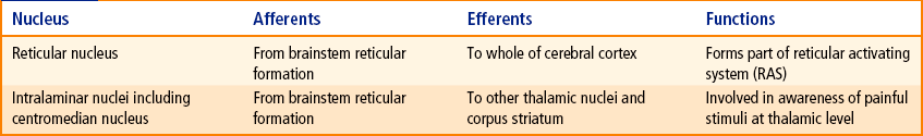
Other thalamic nuclei
Part
Nuclei
Anterior part
Anterior nucleus
Medial part
Medial dorsal nucleus, medial ventral nucleus
Lateral part
• Dorsal tier nuclei
Lateral dorsal, lateral posterior, pulvinar
• Ventral tier nuclei
Ventral anterior (VA), ventral lateral (VL), ventral posterior—(a) ventral posterolateral (VPL), (b) ventral posteromedial (VPM)
Other parts
Intralaminar nuclei, reticular nucleus, medial and lateral geniculate bodies
Connections of thalamic nuclei (Figs 11.4, 11.5)
Connections of the specific nuclei
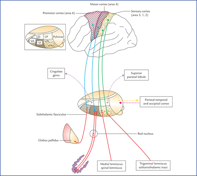
Connections of the nonspecific nuclei
Functions of thalamus
Metathalamus
![]()
Stay updated, free articles. Join our Telegram channel

Full access? Get Clinical Tree


Diencephalon and third ventricle