Fig. 6.1
Peripheral polyneuropathy distribution
In many patients with diabetic polyneuropathy, damage to small axons leads to persistent foot pain (see chapter 20 on pain). The pain, typically described as burning, constant, prickling, and painful to light touch (allodynia) , may be so uncomfortable that the patient seeks medical attention. This pain may be more severe at night and cause disruptions in sleep .
Large, sensory, myelinated axon damage with subsequent loss of vibration and position sense in toes and feet produces gait and balance problems. Motor nerve axons may be involved with weakness of intrinsic foot muscles.
Autonomic nerve axons are also impaired, leading to loss of sweating, thinning of involved skin, asymmetrical pupils that poorly accommodate to darkness, erectile dysfunction, loss of ejaculation, constipation and/or diarrhea, and orthostatic hypotension. Other common manifestations of the autonomic neuropathy in diabetes are resting tachycardia and gastroparesis.
Major Laboratory Findings
Since the neuropathy begins distally in the feet and involves unmyelinated axons, nerve conduction studies of sensory and motor nerves of the leg may initially show mild changes as electrophysiological studies seldom detect abnormalities in unmyelinated axons. With more symptoms developing, nerve conduction velocities slow due to demyelination and loss of myelinated fibers. As the neuropathy progresses, the findings of axonal degeneration predominate with diminished amplitude of compound muscle action potentials and sensory nerve action potentials—due to nerve fiber loss. Needle electromyography of intrinsic foot muscles shows denervation potentials. There is relative preservation of proximal conduction velocities.
A nerve biopsy, while seldom performed, shows non-specific axonal damage to both myelinated and unmyelinated axons. A nerve biopsy should come from a sensory nerve (like the sural nerve) that has a small area of sensory innervation. Biopsy of a mixed nerve will lead to paralysis of innervated muscles and thus is only performed on an intercostal nerve.
Skin punch biopsy (3–4 mm full thickness biopsy) with immunohistochemical staining for peripheral nerve axons is becoming more popular. The histologic sections demonstrate marked reduction in the density of epidermal nerve fibers that is helpful, but not diagnostic, for diabetic neuropathy. Thus, the diagnosis relies mainly on the clinical history, neurologic examination of the peripheral nervous system , and exclusion of other etiologies. However, when a peripheral nerve is affected by two different diseases, the signs and symptoms of a peripheral neuropathy develop earlier, produce more intense symptoms, and have a poorer recovery.
Principles of Management and Prognosis
The management of diabetic neuropathy can be divided into preventing progression of the neuropathy, minimizing problems from anesthesia of feet and hands, and reducing the burning foot pain.
Numerous studies demonstrate that good control of blood glucose can slow, halt, or reverse progression of the neuropathy. Glucose control involves weight loss, exercise, and use of hypoglycemic agents. While optimum glycemic control can reduce the risk of neuropathy, the risk of transient hypoglycemia increases.
Foot anesthesia predisposes to ulceration and infection. Proper footwear minimizes foot and ankle trauma. The individual should be instructed to regularly inspect their feet for signs of infection or ulceration and to place their hand in shoes to detect objects in the soles. If loss of position sense in the feet declines, the patient should use night-lights and caution when walking on uneven surfaces or in the dark.
Treating the patient with a painful peripheral neuropathy is a challenge. For many patients, the pain is reduced with tricyclic antidepressants (amitriptyline and nortriptyline) in low doses. Tricyclic antidepressant medications occasionally can increase the effects of postural hypotension in patients with autonomic neuropathy. Anticonvulsants, such as gabapentin or carbamazepine, may be slowly added if the pain relief is insufficient. In some patients, the foot pain spontaneously subsides when the sensory neuropathy progresses to anesthesia.
Carpal Tunnel Syndrome
Introduction
Carpal tunnel syndrome (CTS) is an example of compression mononeuropathy. Up to 15 % of individuals experience occasional symptoms suggestive of CTS. However, the prevalence of symptomatic carpal tunnel syndrome is estimated at up to 9.2 % in women and 6 % in men with the peak prevalence in older women. CTS can be seen in work-related musculoskeletal disorders caused by repetitive movements and strain. Fortunately, few individuals develop sufficient signs and symptoms to require surgical treatment.
Pathophysiology
The pathophysiology of CTS is not completely worked out but the final step is compression of the median nerve in the carpal canal under the transverse carpal ligament. Several mechanisms may be involved including lesions reducing the size of the carpal tunnel, processes causing swelling of the tendon sheaths such as overuse, and tissue swelling from fluid retention as in pregnancy or myxoedema. MRI findings include swelling of the median nerve just proximal to the carpal tunnel, flattening of the nerve within the carpal tunnel, bowing of the flexor retinaculum, and increased signal intensity of the median nerve. With the addition of gadolinium, the MRI often demonstrates intraneural edema in the median nerve.
About one-third of patients have associated conditions such as pregnancy, inflammatory arthritis, Colles’ fracture, amyloidosis, hypothyroidism, diabetes mellitus, acromegaly, wrist infections, obesity, alcoholism , malnutrition, and use of corticosteroids or estrogens. The remaining two-thirds have their CTS associated with repetitive, often forceful, activities of the hand and wrist.
Major Clinical Features
The symptoms and signs of CTS correspond to the distribution of the distal median nerve (Fig. 6.2b). Patients usually complain of pain, tingling, burning, and numbness that involve the palmar aspect of the thumb, index finger, middle finger, and often the radial half of the ring finger. The fifth digit is only occasionally involved. The symptoms, often worse at night, may awaken the individual with hand discomfort extending into the lower arm that causes the individual to shake their hand (“flick sign”). Symptoms tend to be worse following a day of increased repetitive activity and often increase with driving.
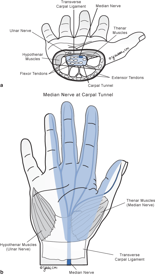
Fig. 6.2


Median nerve. a Carpal tunnel syndrome. b Sensory distribution
Related posts:


Stay updated, free articles. Join our Telegram channel
Full access? Get Clinical Tree
