Fig. 5.1
In myasthenia gravis, acetylcholine receptor antibody blocks the acetylcholine-binding site

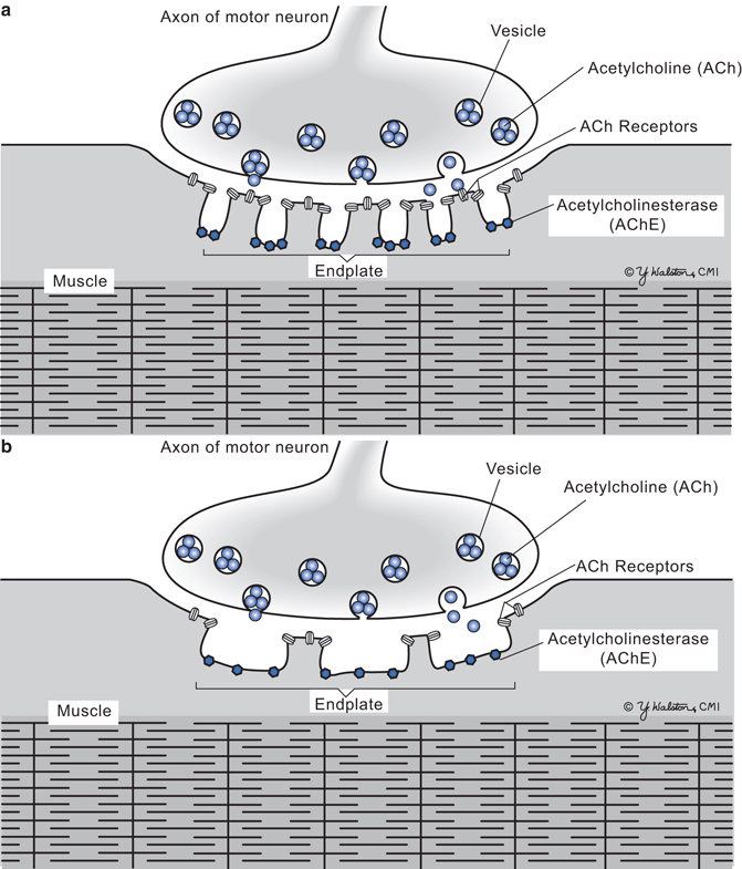
Fig. 5.2
Neuromuscular junction. a Normal. b Myasthenia gravis with simplified post-synaptic membrane
The majority of early-onset, AchR + patients have an associated abnormality of their thymus gland. About 65 % of these patients have thymic hyperplasia with germinal center lymphocyte proliferation, and 10 % have a thymoma. Within the thymus, myoid cells (striated muscle cells) express AchR and may play a role in “priming” helper T cells within the thymus. It is not clear what might be the trigger for the development of the autoimmune response seen in MG, but the elements for the generation and maintenance of autoimmunity exist within the thymus. Surgical removal of the thymus gland often results in clinical improvement and a reduction in the number of circulating antibodies. In older individuals, the thymus is typically atrophic and it is not clear whether it plays a role in the autoimmune response.
Although AchR antibodies are often found, there were many patients who were considered “seronegative” and who did not have AchR antibodies detectable. There are several other antibodies that may play a role in “seronegative” MG, such as muscle-specific kinase (MuSK) antibodies. These are found in up to 70 % of “seronegative” MG patients. MuSK plays a role in clustering of AchR at the NMJ. These patients are more likely to have swallowing and respiratory difficulties at their presentation than AchR+ patients.
Myasthenia gravis can occur in infants. Infants born to mothers with MG may have sufficient circulating antibodies to cause the infant to become floppy, weak, and have a poor suck. This transient syndrome lasts for several weeks until the maternal antibody disappears. Other infants have congenital MG that is due to genetic mutations in the AchR. These infants remain persistently weak and do not respond to immunosuppressive drugs.
Major Clinical Features
The clinical features result from blockade at the neuromuscular junction and affect skeletal muscles in a fluctuating and fatigable manner (Table 5.1). The disease usually has a subacute onset. Earliest symptoms are ptosis and diplopia. Patients complain of droopy eyelids and double vision that will vary during the day and worsen as the day progresses. In 80 % of patients, they will begin with ocular symptoms and then progress to generalized weakness. Signs of bulbar muscle weakness appear with trouble chewing, swallowing, and speaking loudly—although for a minority of patients, the bulbar symptoms will be prominent early in the disease course. Some patients find they eat their big “dinner” meal for breakfast as they have trouble chewing meat by the end of the day. Limb weakness is common and affects proximal muscles greater than distal muscles. Although brief maximal muscle testing may appear normal, patients often cannot hold their arms outstretched for even a minute without fatigue. In severe cases, patients cannot walk and develop respiratory weakness. Sensation, mentation, and deep tendon reflexes are not affected.
Table 5.1
Cardinal features of myasthenia gravis
Weakness | Bulbar muscles: ptosis, diplopia, dysarthia, dysphagia, chewing difficulty Limb muscles: proximal > distal |
Fatigability of skeletal muscles Normal | Increased weakness in afternoon or after exercise Mentation Sensation Deep tendon reflexes |
Maximal weakness appears within the first 3 years of clinical onset. About 10 % of patients experience a spontaneous remission that occurs within the first 2 years. However, the rest of patients have a lifelong chronic illness that fluctuates in severity.
Major Laboratory Findings
Serum antibodies directed against the AchR are found in over 85 % of patients with generalized MG. MuSK antibodies may be detected in patients with “seronegative” MG. The level of antibody titer does not always reflect disease severity as the test detects all AchR antibodies including those that do not interfere with the functioning of the channel. However, for a given patient, a falling titer does reflect clinical improvement.
X-ray or CT of chest may demonstrate a thymoma. Elevated thyroxin blood levels indicating thyrotoxicosis are found in up to 5 %.
Repetitive nerve stimulation (at rate of 3 per second) of proximal muscles (often the trapezius muscle) usually demonstrates a decremental fall of greater than 15 % in the compound muscle action potential (see chapter 3 on common neurologic tests).
Tensilon test: This office test is helpful to establish the diagnosis of MG when there are clear ocular signs—although is performed rarely. Edrophonium (Tensilon) is a brief-acting anticholinergic drug that is slowly given intravenously to a patient. For the next 5–10 min, an untreated MG patient should have a clear objective improvement in ptosis. Often a saline injection precedes the administration of edrophonium to evaluate for a placebo effect.
More commonly, application of crushed ice in a latex glove to a patient’s eye and looking for improvement in ptosis can be performed in the office. This test has a sensitivity of 90 % in distinguishing ptosis due to MG from other causes.
Principles of Management and Prognosis
The goal of treatment is to improve strength and to reduce or eliminate circulating antibodies against the AchR. Symptomatic treatment aimed at improving strength is accomplished with anticholinesterase drugs. These drugs do not reduce circulating antibody titers but are the first line to improve the patient’s strength. Pyridostigmine (Mestinon) is the main oral drug that is given to the patient several times a day. Anticholinergic medications act by interfering with acetylcholine esterase, the enzyme that cleaves acetylcholine in the synaptic cleft. Partial inhibition of this enzyme results in a longer time period that acetylcholine molecules can remain in the synaptic cleft to find unblocked AchR and increase the probability that sufficient AchR channels will open to fully depolarize and contract the muscle fiber. Too much pyridostigmine, however, can block all the acetylcholine esterase such that acetylcholine cannot be cleaved and removed once it attaches to an AchR. The inability to remove acetylcholine from the receptor causes a depolarizing muscle weakness that is called a “cholinergic crisis”. In addition to weakness, a cholinergic crisis is characterized by hyperhidrosis, salivation, and lacrimation.
A number of treatments are aimed at reducing the amount of circulation antibody. Thymectomy, the surgical removal of the thymus gland, in a moderately severe patient often results in clinical improvement and a fall in antibody titer. Corticosteroids and other immunosuppressive drugs (azathioprine and cyclosporine) are commonly given to lower the antibody titer and improve strength. Recently, mycophenolate mofetil has been widely adopted in the treatment of MG due to its minimal side effect profile, but efficacy is somewhat controversial at this point. Intravenous immune globulin (IVIg) and plasma exchange by plasmaphoresis will temporarily reduce circulating antibody and improve strength for several weeks. These temporary methods can be used for patients requiring prompt clinical improvement such as for elective surgery, pneumonia, or a “myasthenic crisis”.
Stay updated, free articles. Join our Telegram channel

Full access? Get Clinical Tree


