9 This chapter discusses the use of embolization in the treatment of spinal neoplasms. To decide whether embolization is indicated, treating physicians must have a clear understanding of the overall treatment plan and the capabilities of endovascular therapy so that realistic goals can be set and achieved with acceptable rates of morbidity. In the early 1960s Djindjian and Di Chiro introduced selective spinal angiography, but applications to spinal embolization were limited until improved devices became available in the 1970s and 1980s. The most important developments were variable-stiffness microcatheters and suitable embolic materials. The former are catheters that are stiffer at their proximal end than at their distal end. This construction creates a very small, soft tip that can be navigated further distally, and therefore into smaller vessels than was previously possible. Such microcatheters permitted percutaneous vascular access to areas that once could only be reached through direct surgical exploration. Access to areas that may not have even been accessible surgically also became possible. In turn, it became feasible to deliver embolic agents in a superselective manner. In the less than 20 years since the introduction of high-quality, readily available, variable-stiffness microcatheters, both technology and techniques have evolved rapidly. These advances have enabled embolization to play a more prominent role in the treatment of neurologic conditions. The spinal cord receives its nourishment from one anterior spinal artery (ASA) and two posterior spinal arteries. The ASA lies in the anterior median sulcus, and the paired posterior spinal arteries lie on either side of the dorsolateral surface of the spinal cord. The numerous branches of the ASA travel within the anterior median sulcus and are responsible for more than two thirds of the blood supply to the spinal cord, including to the anterior and lateral corticospinal tracts. The posterior spinal arteries primarily supply the posterior columns. The paired ASAs originate at the craniovertebral junction as branches of the fourth segment of the vertebral artery. The ASAs arise distal to the origin of the posterior inferior cerebellar arteries. The two ASAs then converge to form a single ASA ventral to the caudal brainstem and rostral spinal cord. The ASA continues within the anterior median sulcus the entire extent of the spinal cord and conus medullaris. In the cervical region, the ASA receives further contributions from branches exiting the vertebral artery and from the ascending cervical branch of the thyrocervical trunk. One anatomically constant radicular artery at C5 or C6 is known as the artery of cervical enlargement. In the thoracic and lumbar regions, radiculomedullary branches of the supreme intercostal arteries and the thoracic and lumbar radicular arteries supply the ASA. The most prominent of these radicular vessels is known as the arterial radicularis magna, or the artery of Adamkiewicz. This artery typically arises from the lower intercostal or lumbar artery on the left side between T10 and L2. It is a slender, midline vessel with a diameter between 0.5 and 1.0 mm. The artery of Adamkiewicz must be identified angiographically before middle to lower thoracic and rostral lumbar lesions are embolized. On an anteroposterior projection, the artery has an ascending segment that makes a characteristic hairpin turn into a descending ASA located in the midline. A pair of posterior spinal arteries occupies the posterolateral aspect of the spinal cord. They originate in the upper cervical region, typically branching from the vertebral artery. They are supplied by the segmental radiculomedullary arteries, which form a hairpin configuration similar to the supply of the ASA. The ASA anastomoses with the posterior spinal arteries at the conus medullaris to form a luxuriant arterial network. The anterior and lateral aspects of the cervical vertebral bodies are primarily supplied by branches of the vertebral artery and the ascending cervical branch of the thyrocervical trunk. In the thoracic and lumbar spine, branches of the lumbar radicular arteries or the intercostal arteries predominantly provide the blood supply to the vertebral bodies. The epidural space ventral and dorsal to the spinal cord and thecal sac contains a rich plexus of vessels. In the ventral epidural space, these vessels lie beneath the posterior longitudinal ligament and contribute to the vascularity of the vertebral body. In the dorsal epidural space, these vessels richly supply the lamina and a portion of the posterior spinous process. A plexus, formed primarily by the main trunk of the dorsispinal artery, lines the outer surface of the lamina and the posterior spinous process. The ability to perform high-quality spinal angiography is a prerequisite for embolization of spinal lesions. Highresolution, preferably biplanar, angiography is required. Although optional, we prefer to place patients under general anesthesia, which minimizes motion and improves image quality, for all but the most straightforward embolization procedures. Patient motion degrades image quality because subtraction techniques are used to provide detailed images. For subtraction techniques, a mask image must first be acquired. After contrast is injected, the original mask image is subtracted from the subsequent images, greatly enhancing visibility of the contrast agent. Patient motion between the time of the initial or mask image and the time of the contrast-injected images markedly degrades image quality. The importance of minimizing this motion artifact becomes even greater when interventions are undertaken. For example, there must be no patient motion during the injection of an embolic agent, or the embolic material will be poorly visualized. Similarly, when catheters and endovascular devices are manipulated during procedures, progress is monitored visually using real-time subtracted fluoroscopic imaging known as “road maps.” As long as there is no motion, these images remain clear. Even subtle movements, however, markedly degrade image quality. Motion from breathing and even intestinal peristaltic movements can render images un-interpretable. Glucagon can be administered to reduce intestinal motility, again helping to minimize motion artifact and to enhance image quality. Likewise, ventilation can be suspended during key periods of image acquisition or during delivery of embolic agents. During the process of optimizing image quality, it is equally important that patients and staff be protected from excessive exposure to radiation. Appropriate attention must be paid to radiation shielding, the length of fluoroscopy, and other issues related to radiation exposure. When endovascular neurosurgical procedures are performed with the patient under general anesthesia, many surgeons advocate the use of electrophysiologic monitoring to compensate for the lack of clinical monitoring. Somatosensory evoked potentials are a sensitive measure of spinal cord function and have become a requisite adjunct during spinal endovascular procedures.1–4 Anecdotally, monitoring of motor evoked potentials has been reported to be particularly useful.5 Microcatheters are categorized as one of two types: over the wire or flow directed. Over-the-wire systems are advanced toward their target using a fine, curved guidewire that is steered toward the target area using torque to direct the wire. The catheter is advanced over the wire. Flow-directed catheters tend to be softer than over-the-wire catheters. Their distal tips are carried passively with arterial blood flow. Because these catheters depend on blood flow, they are best suited to high-flow lesions such as arteriovenous malformations (AVMs). Hybrid catheters that can be directed by blood flow but with some wire assistance are now being produced. Many variations are available within each class of catheter. Embolic agents are classified as liquids or particulates (Table 9-1). Liquid agents may be glue-type agents such as N-butyl-cyanoacrylate (NBCA) or sclerosing agents such as ethanol. Recently, Onyx™ liquid embolic system (Micro Therapeutics, Irvine, CA) has been used to treat spinal AVMs with good results. Particulate agents may be larger particles such as metallic coils (generally platinum) or smaller injected particles such as polyvinyl alcohol (PVA). In general the larger a particle is, the more proximal is the vascular occlusion in relation to the target lesion. An alternative method of devascularizing hypervascular tumors involves preoperative percutaneous intralesional injection of alcohol agents.6 This modality may be useful if the spinal cord and tumor share the same vascular supply, a situation that precludes transarterial embolization. Ethanol can cause tumor necrosis and may facilitate surgical excision. Symptoms have improved after the percutaneous delivery of ethanol in vertebral hemangiomas.7 However, osteonecrosis of the affected vertebral body may cause a compression fracture.6,7
Embolization Techniques for Neoplasms of the Spine and Spinal Cord
 Spinal Vascular Anatomy
Spinal Cord
Anterior Spinal Artery
Posterior Spinal Artery
Spinal Column
 Endovascular Techniques
Microcatheters
Embolic Agents
Percutaneous Embolization
| Liquids N-butyl cyanoacrylate (NBCA) Onyx Ethanol Fibrin glue Particulates Polyvinyl alcohol (PVA) Cellulose beads and microspheres Microfibrillar collagen Gelatin foam Metallic coils |
 Rationale for Embolization of Spinal Neoplasms
There is obvious appeal in the idea of devascularizing hypervascular tumors before they are resected surgically. Predictably, certain tumors, particularly hemangioblastomas and metastatic tumors such as renal and thyroid metastases, are highly vascular (Table 9-2). Consequently, they may be difficult to resect.
Embolization techniques can be divided into transarterial, direct puncture, and intraoperative. Although transarterial embolization of spinal column tumors has become a widely accepted adjunct to surgical resection, the literature supporting this approach is limited. Case series have described good results associated with minimal morbidity.8–11 However, case series tend to be small, uncontrolled, and retrospective and usually include heterogeneous types of tumor. Some authors have attempted a more systematic evaluation. Gellad et al12 reviewed 24 patients with spinal metastases and compared blood loss in those whose tumors were adequately embolized with that in those whose embolization was inadequate or not attempted. During resection blood loss for the two groups averaged 1850 and 3500 mL, respectively. Hess and co-workers10 studied 17 patients with spinal metastases and also found that the mean blood loss at tumor resection was significantly less after embolization (2088 mL after embolization compared with 3500 mL without embolization). The reference group was matched with respect to tumor pathology, location, and surgical procedure.
| Primary Benign Hemangiomas Aneurysmal bone cysts Osteoblastomas Malignant Giant cell tumors Chordomas Plasmacytomas Osteogenic sarcomas Chondrosarcomas Epithelioid hemangioendotheliomas Metastatic Renal cell carcinomas Thyroid carcinomas Hepatocellular carcinomas Neuroendocrine tumors Germ cell tumors |
Manke and co-workers13 studied a more homogeneous group of patients—only those treated for renal metastases. They compared 20 metastases (in 17 patients) that were embolized with 11 metastases (in 10 patients) that were not embolized. Embolized patients were subdivided into two groups depending on the thoroughness of the embolization procedure. Surgical blood loss was significantly less in patients receiving preoperative embolization even if the embolization was only partial. The mean blood loss for no embolization, partial embolization, and complete embolization was 5000, 2000, and 1500 mL, respectively. Olerud et al14 evaluated 21 patients with metastatic thoracolumbar renal cell carcinomas. In patients who had undergone preoperative embolization, blood loss during surgery was less than in patients who had not been embolized regardless of approach. No complications were associated with embolization (a total of 11 procedures).
A legitimate question is whether this reduction in blood loss is clinically important. In addition to the objective decrease in blood loss, most authors subjectively report that complete tumor resection is more readily accomplished after embolization.10,15 Although it seems probable that embolization facilitates tumor resection, it is difficult to find data that demonstrate improved patient outcomes. At present the most objective evaluation of the value of preoperative tumor embolization remains the indirect marker of efficacy of decreased surgical blood loss.
Few studies have attempted a systematic comparison of embolization techniques. Berkefeld and co-workers16 correlated embolic agents to intraoperative blood loss in 59 patients who underwent vertebral corpectomies. Ten patients did not undergo embolization, and their mean operative blood loss was 4350 mL. This finding was not significantly different from that of patients who underwent coil embolization of tumor feeding arteries in whom the mean blood loss was 2650 mL. In contrast, patients who underwent embolization with PVA, which penetrates more distally into a tumor, had a statistically significant lower mean blood loss of 1800 mL.
Proximal feeding artery occlusions are considered less helpful than embolization procedures that deliver the embolic agent into the tumor itself. The latter technique reduces the possibility that collateral circulation will continue to supply the tumor despite occlusion of the proximal feeding artery. Manke and co-workers13 evaluated embolization for renal cell carcinoma metastatic to the spine by correlating the particle size of PVA used with the amount of intraoperative blood loss. There was no significant difference in blood loss when particles were smaller or larger than 250 μm. These studies, however, may be too small to provide reliable comparisons of embolization techniques. It is also likely that embolization can be achieved more safely when catheters are positioned within the tumor vessels themselves, thereby reducing the chance of embolizing other vessels.
 Intramedullary Spinal Neoplasms
Extensive spinal angiography is necessary for patients with intramedullary tumors frequently requiring examination of several levels above and below the lesion.1 The most common intramedullary spinal tumors include ependymomas, astrocytomas, and hemangioblastomas. Hemangioblastomas are the most hypervascular of these spinal cord tumors. They can occur sporadically as a singular lesion or with multiple central nervous system tumors as a manifestation of the von Hippel-Lindau syndrome. Hemangioblastomas can have robust arterial feeders and typically have a single draining vein, mimicking an intramedullary AVM. If the posterior spinal artery is catheterized superselectively, embolization with a liquid embolic agent may be performed safely.
Reports indicate that preoperative embolization of hemangioblastomas may help mitigate intraoperative blood loss. Tampieri et al17 treated two patients with large hemangioblastomas, one spinal and one involving the posterior fossa, with preoperative embolization. Both lesions were then resected with less than 100 cc of blood loss. Eskridge et al18 treated nine patients with craniospinal hemangioblastomas with polyvinyl alcohol embolisate and incurred no permanent complications. The surgeons believed that embolization facilitated tumor manipulation and surgical resection. Conway et al19 described 4 of 40 patients with hemangioblastomas who underwent preoperative embolization. In a patient with a sacral hemangioblastoma, embolization alone was sufficient to arrest progression of the patient’s symptoms. Lee et al20 treated four of 14 patients with spinal cord hemangioblastomas with embolization and surgical resection. Complete resection was achieved in all four cases. Subjectively, the surgeon reported significantly less blood loss in the embolized patients. Contraindications to embolization included supply to the hemangioblastoma from the artery of Adamkiewicz and a surgically accessible arterial supply. Four patients were treated before the advent of spinal embolization techniques. Tumor feeders emanating from the anterior spinal axis should not be embolized because of the risk of occluding the ASA. Preoperative embolization is unwarranted in patients with an ependymoma or astrocytoma.
 Spinal Column Tumors
The angiographic appearance of spinal tumors is nonspecific. Computed tomography (CT) and magnetic resonance imaging (MRI) findings are far more suggestive of their diagnosis than angiograms. Hemangiomas, however, are notable exceptions in that their appearance on CT, MRI, and angiography is almost pathognomonic. The presence of irregular vessels, rapid staining during the arterial phase, and arteriovenous shunting on angiography suggest malignancy.
For patients with primary or metastatic bone tumors, angiography should encompass two levels above and two levels below the lesion. Large-caliber feeding vessels to the tumor should be embolized systematically. To reduce the risk of early recanalization, a platinum coil may be placed after embolization of a feeder with glue or PVA.
Primary Bone Tumors
Vertebral hemangiomas are the most common benign spinal tumor. Based on large autopsy studies, their incidence is 10 to 12%.21 In most patients, hemangiomas remain asymptomatic. In rare cases, however, hemangiomas may extend into the epidural space, collapse the vertebral body, or compress the thecal sac. When surgical excision is planned for decompression and fixation, angiography and preoperative embolization should be considered. Angiography helps demonstrate the vascularity of the intra- and extraosseous portions of the tumor, identifies the feeding arteries, and delineates the relationship of the vascular supply to the spinal cord. The arterial supply should be embolized if there is no shared supply with the ASA. Many reports and retrospective case series have described successful embolization of these lesions.22–25
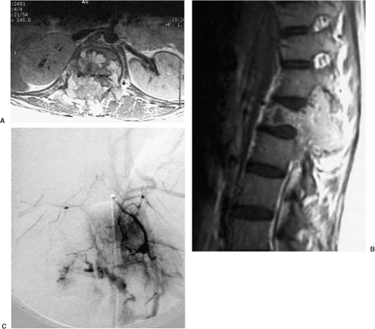
Aneurysmal bone cysts are benign hemorrhagic mass lesions that represent 1% of all primary bone tumors (Fig. 9-1). Their cell of origin is unknown,26–28 and their clinical course is unpredictable. Surgical excision, radiotherapy, and embolization have all been advocated for the treatment of these lesions. Boriani et al29 and De Cristofaro et al30 recommend embolization as the sole treatment for patients without neurologic deficits or spinal instability when their CT or MRI studies are unequivocal. Patients with neurologic deficit, spinal instability, or known recurrence or progression of disease despite previous treatment should undergo surgical excision.
Hemangiopericytomas are rare tumors that originate from pericytes surrounding the reticular sheath of capillaries and postcapillary venules. They most often occur in the lower extremities. Spinal hemangiopericytomas, whether primary or metastatic, are exceptionally rare. Surgical excision is the treatment of choice but is frequently complicated by the invasiveness of the tumor and its hypervascularity. Preoperative angiography helps delineate the vascular supply, and embolization reduces the vascularity of the tumor.31–33 Muraszko et al32 warn that neurologic deterioration related to tumor swelling can immediately follow embolization, necessitating urgent excision of the tumor.
Brennan et al34 described an epithelioid hemangioendothelioma of the cervical spine treated with embolization and subsequent staged resection. These lesions are tumors of vascular origin with rare spinal involvement. Their robust vascularity makes preoperative embolization imperative.
Giant cell tumors compose 4.2% of all primary bone tumors and typically occur in the third decade of life. The sacrum is the most common spinal location for this tumor (Fig. 9-2). Surgical resection can be fraught with complications, and sacral giant cell tumors are associated with a high rate of local recurrence (~33%).35 Lin et al36 treated 18 patients with giant cell tumors of the sacrum with embolization with or without chemotherapy. In 14 patients pain and neurologic symptoms improved immediately, with durable improvement in about half of the patients (median follow-up, 105 months). One patient died the day after embolization from undetermined causes. The authors recommended that embolization for these tumors be used alone or in conjunction with other treatment modalities.
Spinal Metastatic Lesions
Spinal metastases occur in 5 to 10% of cancer patients.37 The most common metastatic lesions to the spine are breast, prostate, and lung carcinomas. Spinal metastases have implications for both quality of life and survivability. Metastases can cause intractable pain. They can impair motor function, which is functionally devastating, and can predispose patients to medical complications related to immobility (e.g., deep venous thrombosis, pulmonary emboli, pneumonia); therefore, surgical decompression and resection play a prominent role in the management of these patients.
Known hypervascular metastatic tumors include renal cell carcinomas, thyroid carcinomas, melanomas, sarcomas, and neuroendocrine tumors. These lesions often affect the thoracic spine and extensively involve the epidural and paraspinal spaces. Preoperative embolization should be strongly considered if the lesion is thought to be hypervascular.
Magnetic resonance imaging can predict the degree of vascularity of spinal tumors with modest sensitivity and specificity. MRI features suggestive of hypervascularity include large flow voids within the tumor, bright contrast enhancement, and signs of hemorrhage on T1- and T2-weighted images. Prabhu et al15 graded the vascularity of 51 metastatic spinal tumors on angiography and MRI and correlated the two studies. The positive predictive value of MRI was 77%, whereas the negative predictive value was far less (21%).
Related posts:
 Lumbosacral and Pelvic Reconstruction
 Anatomy of the Spine and Spinal Cord
Stay updated, free articles. Join our Telegram channel

Full access? Get Clinical Tree


