Fig. 19.1
Sagittal T1 MRI thoracic spine with contrast demonstrating severe kyphotic deformity at T6/7 due to infection. The kyphosis, along with epidural enhancing tissue, is encroaching upon the spinal cord
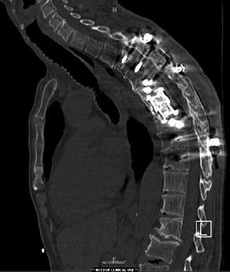
Fig. 19.2
CT scan of the thoracic spine postoperatively demonstrating reconstruction of anterior and middle column via an expandable thoracic cage from a lateral position with elimination of thoracic kyphotic deformity. Second stage of surgery involved posterior instrumentation and fusion
19.3 Conservative Management and Treatment of Thoracic Kyphotic Deformity
Patients without significant vertebral body collapse who are asymptomatic or with minimal pain can be managed conservatively. Conservative management generally involves supervised physical and occupational therapy, bracing with thoracolumbar orthoses for comfort, and anti-inflammatory or narcotic medications that are supervised by a pain management specialist. Additionally, close follow-up of these patients is indicated with upright x-rays assessing progression of thoracic kyphotic deformity that may necessitate movement away from conservative management and toward a surgical path [7, 8].
19.4 Indications and Goals for Surgical Correction of Thoracic Kyphotic Deformity
Indications for surgical correction of spinal deformity include instability, deformity, intractable pain, and current or impending neurological compromise [3].
19.5 Surgical Approaches to Treating Thoracic Kyphotic Deformity
A variety of surgical approaches have been studied for correction of thoracic kyphotic deformity with placement of expandable cages. These include open approaches as well as the more recent minimally invasive techniques utilizing percutaneous instrumentation and endoscopic assistance. The goals of kyphotic deformity correction center around altering the main vector of forces drawing the thoracic spine into the kyphotic position. This is mainly done via reconstructing the anterior and middle columns from a variety of different approaches through the use of expandable thoracic cages.
19.5.1 Posterior
19.5.1.1 Laminectomy/Posterolateral Instrumentation/Osteotomy/Fusion
Posterior techniques for ventral thoracic and thoracolumbar pathology have evolved over the years. Laminectomy with Smith-Petersen osteotomies, along with pedicle subtraction osteotomies, has been shown to improve lordosis approximately 6–10° and 15–20°, respectively, via shortening of the posterior elements [9]. However, these techniques are associated with decreased vertebral height and buckling of the posterior spinal ligaments and dura with the possibility of associated cord compression [10]. Additionally, these techniques are associated with significant blood loss and pulmonary complications [11, 12].
The use of long-segment Harrington rod instrumentation may be used to restore thoracic curvature. However, this technique is fraught with morbidity and complications due to the long-segment fusion, possibility of instrumentation failure requiring reoperation, inability to restore the rotational deformity, and possibility of further worsening the preexisting kyphosis upon failure [13]. Additionally, purely posterior pedicle screw instrumentation with fusion may not be able to withstand the physiologic stress from an anterior vector, resulting in hardware failure and progression of the underlying kyphosis [3, 14]. McLain et al. noted progressively worsening deformity during the first 6 months postoperatively after stand-alone posterior kyphotic reduction maneuvers [14]. Multiple studies have demonstrated a failure rate of 20–50 % with solely posterior pedicular fixation and fusion in patients without anterior support [15–17].
19.5.1.2 Laminectomy/Costotransversectomy with Expandable Cage and Posterolateral Instrumentation/Fusion
Laminectomy with costotransversectomy is a technique that has allowed surgeons to access ventral pathology in the thoracic spine. A unilateral approach with laminectomy and removal of the transverse process and portion of the rib head and proximal rib has allowed access down the pedicle and into the affected vertebral body[s] [3]. This allows placement of a thoracic cage anteriorly via a posterior approach between the exiting nerve roots (usually sacrificed in the thoracic spine allowing ample room) to reconstruct the anterior and middle column. Reconstruction of the anterior and middle columns from this approach is typically reinforced by a short-segment pedicle screw instrumentation and posterolateral fusion [18].
Sciubba et al. describe a novel technique of a purely posterior approach with circumferential costotransversectomy and corpectomy toward treating anterior thoracic pathology [3]. They described performing standard bilateral costotransversectomies with transpedicular corpectomy and placement of expandable thoracic cage. They documented seven cases of circumferential costotransversectomies with placement of expandable thoracic cage and noted a kyphosis improvement of 53 % [3]. They calculated a mean kyphotic angle preoperatively of 28.6° and postoperatively of 12.1° [3]. This effect is in accordance with the so-called boundary effect allowing for a greater surface area of anterior axial loading [19].
Snell et al. have also described a similar approach in 15 patients toward treating thoracic kyphotic deformity [20]. They utilized both expandable and non-expandable thoracic cages for reconstruction and noted adequate neurological stabilization and kyphosis reduction in their cohort with two patients improving at least one Frankel grade [20]. The use of expandable cages allows for appropriate distraction of the thoracic spine and provides an adequate surface area along the superior and inferior end plates to facilitate solid fusion [3]. The use of expandable cages, as opposed to fibular and iliac grafts, decreases complications such as end plate penetration due to the large footprint of the expandable cages [12].
Abumi et al. and Oda et al. described the precise benefit of expandable cages as compared to non-expandable cages during spinal reconstruction [21, 22]. They noted the former have a greater in-line distraction capability of the spinal ligaments, which may improve fusion rates [21]. Additionally, the ability to manually distract while noting expansion both visually and radiographically of vertebral height is quite user-friendly in assuring restoration of lordosis and minimizing kyphotic tendency around the normal internal axis of rotation of the thoracic and thoracolumbar spine [3]. Finally, non-expandable cages require one additional step of posterior compression of instrumentation, whereas use of expandable cages may avoid this process [23]. In fact, Knop et al. studied 12 cadaveric spines and biomechanically found more stabilization using an expandable cage compared to the non-expandable cage and noted a decreased need for posterior compression when the expandable cage was used [23]. An additional prospective study using expandable cages by Lange et al. showed successful stabilization of anterior column with no failures in 126 patients with infection, tumor, and traumatic pathology [24]. This led to the development of a larger-size forceps spreader to increase the height of this expandable cage one more level [24]. Keshavarzi et al. retrospectively studied 35 patients from two large centers with thoracic kyphotic deformity due to infection, trauma, and tumor who underwent corpectomy and placement of expandable thoracic cages. They noted early postoperative correction in kyphosis in all, restoration of sagittal alignment at 12 months, and reduction in visual analog pain scale over the 31-month follow-up period [25].
Overall, this technique avoids the morbidity of a large thoracoabdominal and/or transthoracic exposure while completely decompressing neural structures, stabilizing the anterior and middle columns, and restoring adequate sagittal balance. The autograft obtained from the initial decompression can be utilized within the cage itself, allowing for successful fusion via osteoconductive and osteoinductive properties of stem cells. Lastly, supplementing posterior instrumentation with an anterior expandable cage allows for minimizing hardware failure and potentially decreasing the rate of pseudoarthrosis [26].
19.5.2 Anterolateral
Anterior and anterolateral techniques for thoracolumbar kyphotic treatment include the transthoracic-transpleural thoracotomy, thoracoscopy using endoscopic approaches, and a more standard thoracoabdominal/retropleural approach [5]. These techniques have all been well described and utilized in treating this pathology. Compared to the posterior techniques described above, many claimed that patients with respiratory dysfunction and significant comorbidities often are not candidates for this anterior-anterolateral approach in accessing the anterior thoracic spine [3, 5]. Complications noted via these approaches include persistent pleural effusions, hemothorax, chylothorax, and dural-pleural fistulae [27, 28]. Additionally, these procedures typically will obviate the need for a second stage surgery for posterior pedicle instrumentation and fusion at some point, which increases operative time for the patient as well as morbidity and blood loss [5].
Related posts:

Stay updated, free articles. Join our Telegram channel

Full access? Get Clinical Tree


