Fig. 70.1
Fibrous dysplasia. (a) Coronal T1-weighted precontrast MR image. (b) Coronal T1-weighted gadolinium-enhanced image. (c) Sagittal T1-weighted gadolinium-enhanced image. (d) Axial T1-weighted gadolinium-enhanced image. An expansile lesion is centered in the body of the sphenoid, demonstrating heterogeneous signal intensity and faint contrast enhancement. The pituitary gland is posterior to the lesion

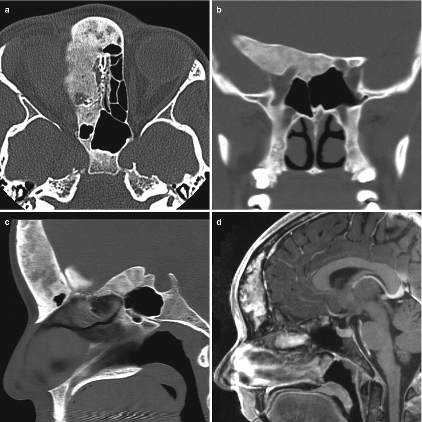
Fig. 70.2
Fibrous dysplasia. (a) Axial CT image in bone algorithm. (b) Coronal CT image in bone window. (c) Sagittal CT image in bone algorithm. (d) Sagittal T1-weighted gadolinium-enhanced MR image. Bony expansion with ground-glass attenuation is observed in the right frontal bone, right ethmoid bone, and right sphenoid wing. The lesion involves the medial right orbital wall and displaces the orbital contents. MRI shows heterogeneous enhancement within the lesion
70.3 Histopathology
FD is characterized by abnormal fibroblast maturation and proliferation resulting in replacement of normal bone with weaker, immature woven bone.
FD rarely undergoes malignant transformation to osteosarcoma, fibrosarcoma, or chondrosarcoma [22].
70.4 Clinical and Surgical Management
Conservative management is recommended in most cases, as the disease is often benign and self-limited.
Corticosteroids may benefit some patients with progressive visual changes [27].
A biopsy may be necessary if the imaging features are atypical or questionable, especially in a monostotic lesion where the diagnosis has not yet been established.
Surgical decompression (especially optic nerve decompression) may be warranted in a minority of cases with cranial nerve compression or craniocervical joint involvement [10, 16].
A transsphenoidal approach may be used to obtain a biopsy specimen or achieve surgical decompression for appropriate skull base lesions [4, 28].
In recent years, extended endoscopic approaches have been used to treat symptomatic FD lesions [4, 29].
In cases of McCune-Albright syndrome, primary management of the endocrinopathy and hypersecretion syndromes using surgery, medications, and/or radiation may be needed to achieve endocrinological remission.
References
1.
Lipson A, Hsu TH. The Albright syndrome associated with acromegaly: report of a case and review of the literature. Johns Hopkins Med J. 1981;149:10–4.PubMed
2.
3.
4.
5.
Christoforidis A, Maniadaki I, Stanhope R. McCune-Albright syndrome: growth hormone and prolactin hypersecretion. J Pediatr Endocrinol Metab. 2006;19:623–5.CrossRefPubMed
Stay updated, free articles. Join our Telegram channel

Full access? Get Clinical Tree


