Fig. 37.1
Germ cell tumor. (a) Sagittal T1-weighted pre-gadolinium image. (b) Sagittal T1-weighted post-gadolinium image. (c, d) Coronal T1-weighted post-gadolinium image. There is a lobular, heterogeneously enhancing mass in the sella and suprasellar regions, with involvement of the pituitary stalk (b, arrow) and the floor of the third ventricle. Additional nodular enhancing lesion is seen in the right lateral ventricle (c, arrowhead), consistent with distant spread of tumor

Fig. 37.2
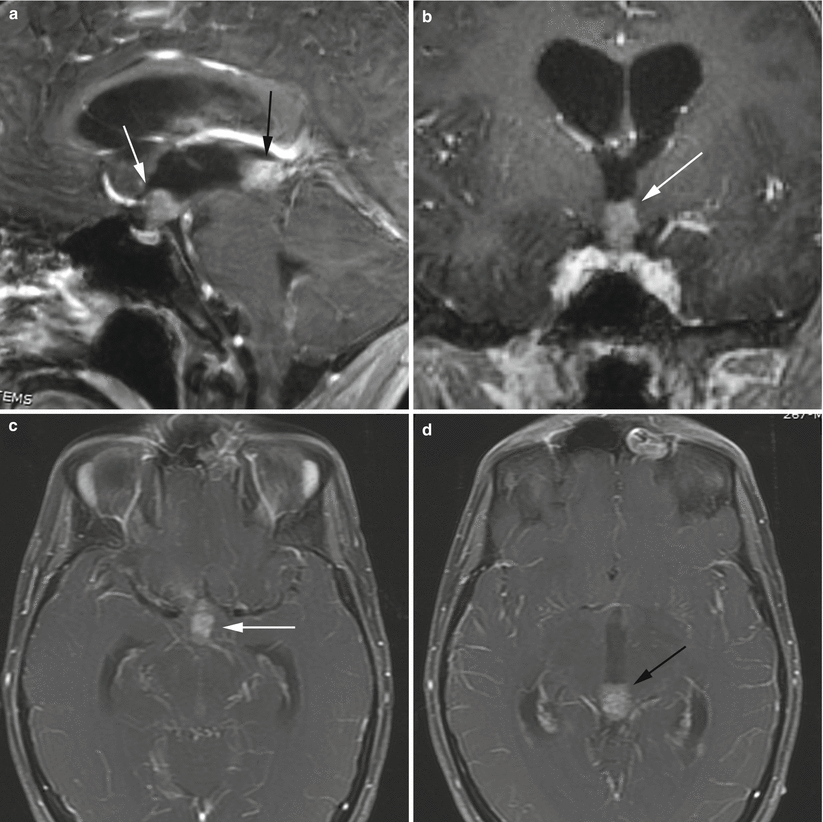
Germinoma. (a) Sagittal T1-weighted post-gadolinium image. (b) Coronal T1-weighted post-gadolinium image. (c, d) Axial T1-weighted, fat-saturated, post-gadolinium image. There are nodular enhancing masses in both the suprasellar region (a, d, white arrows) and the pineal region (a–c, black arrows). The suprasellar lesion involves the pituitary stalk, but the pituitary gland appears separate from the mass. There is hydrocephalus with enlargement of the lateral and third ventricles
37.3 Histopathology
Germ cell tumors of the sellar or suprasellar region are histologically identical to their gonadal and extragonadal counterparts.
The tumors may be divided into two major groups: germinomas and nongerminomatous germ cell tumors. The nongerminomatous group includes embryonal carcinoma, endodermal sinus tumor (yolk-sac tumor), choriocarcinoma, and teratomas (mature, immature, or with malignant transformation) [18].
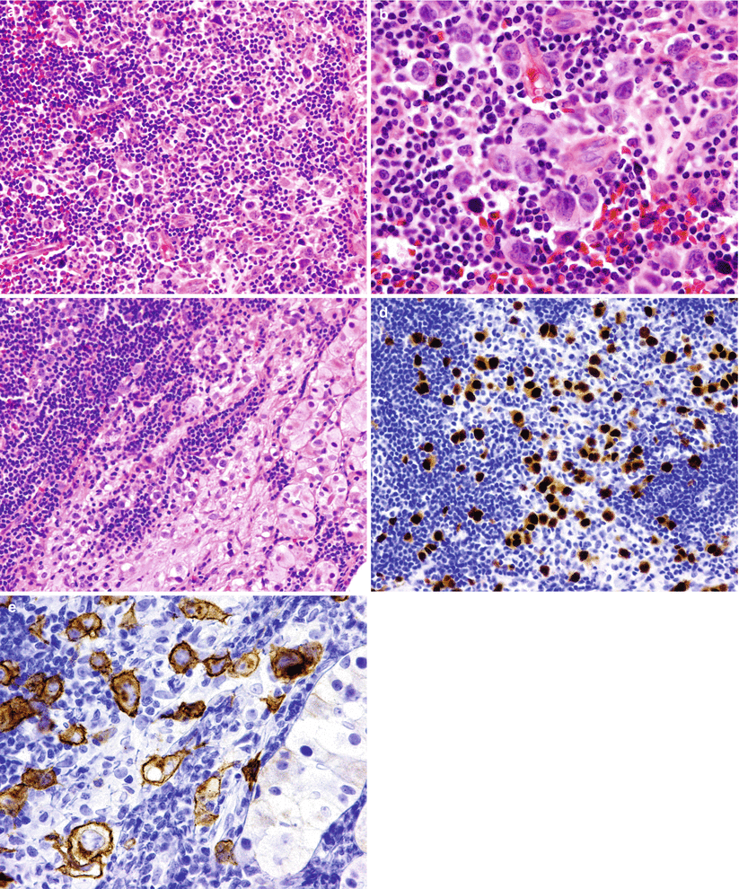
In the sellar/suprasellar region, germinoma (Fig. 37.3) is the most frequent germ cell tumor, accounting for 61.5 % of cases, followed by teratomas and mixed germ cell tumors [19, 20].
In addition to specific histopathologic patterns, several immunomarkers are helpful to differentiate these tumor subtypes (e.g. OCT4, c-kit).
In children presenting with diabetes insipidus and hypopituitarism who undergo surgical biopsy, germ cell tumors may be masked by lymphocytic infiltration and demonstrate a pathology that is initially more consistent with hypophysitis. Delayed development of germ cell tumors frequently follows, however, even up to several years later [21, 22].

Fig. 37.3
Germinoma. An intrasellar germinoma showing large cells with prominent vesicular nuclei intermixed with a rich infiltrate of lymphocytes (a, b). The germinomatous and lymphocytic cells have obliterated the anterior pituitary parenchyma, with only a few remaining normal neuroendocrine acini (c). Immunohistochemical stains for OCT4 (d) and c-kit (e) highlight the neoplastic large cells in contrast to the stromal lymphocytes
37.4 Clinical and Surgical Management
Patients should undergo serum and CSF evaluation for AFP, carcinoembryonic antigen (CEA), and β-hCG.
Multimodal intervention is frequently required and depends on the histological tumor components [23, 24]:
Localized germinomas can be treated with radiation, frequently with good response rates [25].
Disseminated germinomas are typically treated with chemotherapy.
Mature teratoma components are treated with surgical resection.
β-hCG and AFP levels in the serum and CSF may be used to monitor treatment progress.
Prognosis is worse for embryonal carcinomas and choriocarcinomas than for germinomas or mature teratomas [23].
The 5-year survival rate for patients undergoing radiation for germinomas is 85 %, compared with 46 % for patients with nongerminomatous germ cell tumors [6].
References
1.
2.
Nishio S, Inamura T, Takeshita I, Fukui M, Kamikaseda K. Germ cell tumor in the hypothalamo-neurohypophysial region: clinical features and treatment. Neurosurg Rev. 1993;16:221–7.CrossRefPubMed
Stay updated, free articles. Join our Telegram channel

Full access? Get Clinical Tree


