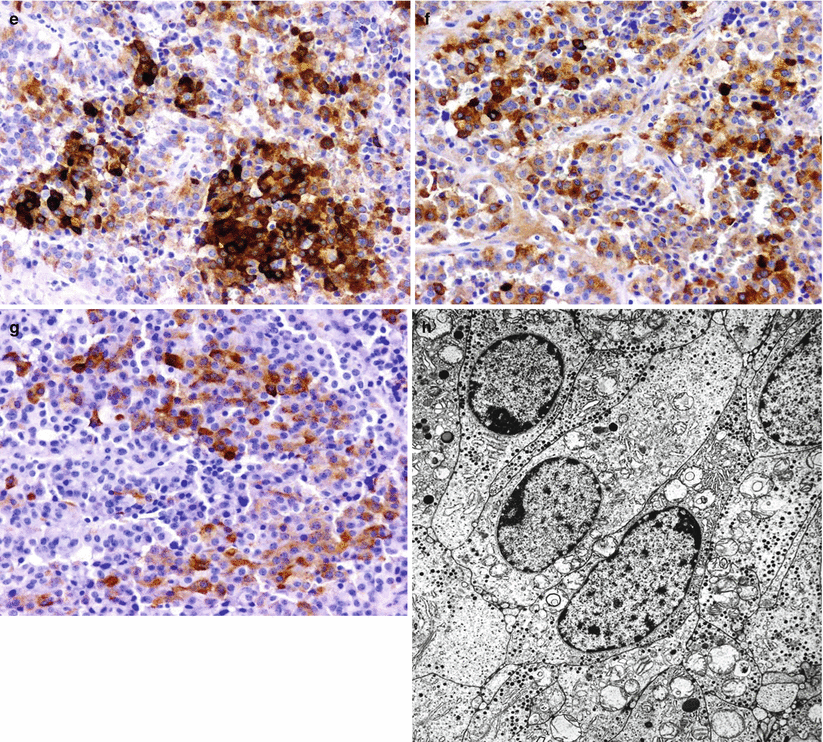
Fig. 16.1
Gonadotropin-secreting adenoma. Gonadotropin-secreting adenomas or gonadotroph cell adenomas are characteristically chromophobic tumors (a) with diverse histopathological arrangements. Tumors may have papillary arrangements (b), nested arrangements (c), and focal oncocytic appearance (d). Gonadotroph adenomas show variable immunoreactivity for follicle-stimulating hormone (FSH) (e), luteinizing hormone (LH) (f), and the alpha-subunit of glycoproteins (g). Gonadotroph cell adenomas are mostly composed of well-differentiated, elongated cells with a degree of cellular polarity at the ultrastructure level. Secretory granules are small and tend to be located at the periphery of the cytoplasm (h)
Electron microscopy may demonstrate different structural features in the adenomas of men versus those of women. Gonadotroph adenomas in men resemble null-cell adenomas, with poorly or moderately developed cytoplasmic organelles. In women, tumors tend to be well differentiated, with distinctive vesicular dilatation of the Golgi complex (“honeycomb Golgi”) [6].
16.3 Clinical Management
As with most functional pituitary adenomas, transsphenoidal surgical resection remains the mainstay of treatment.
Dopamine agonist therapy (cabergoline or bromocriptine) has been reported to be an effective medical treatment for a subset of these tumors [7].
Successful treatment with a gonadotropin-releasing hormone antagonist also has been reported [8].
Resolution of ovarian hyperstimulation, reduction of ovary size, and successful pregnancy have been reported following effective treatment.
References
1.
Young Jr WF, Scheithauer BW, Kovacs KT, Horvath E, Davis DH, Randall RV. Gonadotroph adenoma of the pituitary gland: a clinicopathologic analysis of 100 cases. Mayo Clin Proc. 1996;71:649–56.CrossRefPubMed
Stay updated, free articles. Join our Telegram channel

Full access? Get Clinical Tree


