Fig. 36.1
Granular cell tumor. (a) Coronal T1-weighted pre-gadolinium image. (b) Coronal T1-weighted post-gadolinium image. (c) Sagittal T1-weighted post-gadolinium image. (d) Coronal T2-weighted image. There is a hypoenhancing mass in the suprasellar region anterior to and abutting the pituitary stalk. The mass appears separate from the pituitary gland
36.3 Histopathology
Intraoperatively and grossly, granular cell tumors firm, vascular tumors that may bleed extensively during surgical resection [3].
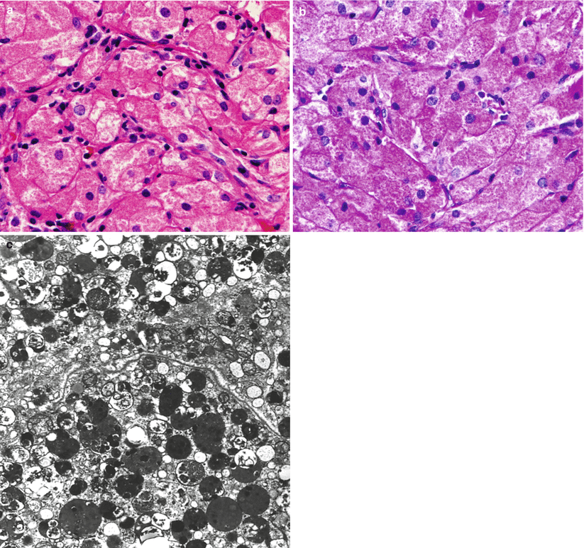
Histologically, granular cell tumors are characterized by uniform polygonal cells with distinct, eosinophilic granular cytoplasm. The nuclei are round with delicate chromatin and uniform nucleoli, and they tend to be located in the periphery of the cells.
The cytoplasmic granules are positive for periodic acid–Schiff (PAS) stain and are diastase resistant (Fig. 36.2) [8].
The tumor shows absent to low mitotic activity, and necrosis is rarely seen.
A fine, delicate network of capillaries is present, with occasional perivascular lymphocytic infiltrates.
Granular cell tumors are negative for neuroendocrine markers.
The tumors show immunoreactivity for vimentin and S100 protein, but only occasionally show GFAP immunoreactivity [8].
The macrophage/lysosome marker CD68 is consistently and strongly immunoreactive in all tumors.
Most significantly, granular cell tumors are strongly immunoreactive for TTF-1, as are the other pituicyte-derived tumors [9, 10].
Electron microscopy shows a typical rich, lysosomal population (Fig. 36.2c). Intermediate filaments are scarce, and neurosecretory granules are absent.

Fig. 36.2


Granular cell tumor. Granular cell tumors show characteristic plump, large cells with granular cytoplasm by H&E stain (a), which is strongly positive by periodic acid–Schiff (PAS) staining after diastase treatment (b). The granular cytoplasmic component is represented by lysosomal accumulation by electron microscopy (c). Similar to other tumors thought to be derived from pituicytes of the neurohypophysis, nuclear immunoreactivity for TTF-1 is present (not shown)
Stay updated, free articles. Join our Telegram channel

Full access? Get Clinical Tree


