Fig. 32.1
Hemangioblastoma. (a, b) T1- and T2-weighted MRI scans taken 2 years prior to presentation. (c) This T1-weighted MRI scan reveals a partly vesicular, hyperintense, and slightly increased (vs a and b) intrasellar and suprasellar mass of 16 mm in diameter, with progressive compression of the prechiasmatic portions of the optic nerves bilaterally. (d) T2-weighted MRI scan showing the vesicular portion as hypointense; normal pituitary tissue could not be clearly delineated. (e, f) No enhancement is evident on T1-weighted imaging after intravenous administration of gadolinium. (g, h) An MRI scan taken 16 months postoperatively showed regular display of the remaining pituitary gland with good chiasmatic decompression and no signs of tumor recurrence (Adapted from Schar et al. [9]; with permission)
32.2 Imaging Features
Infundibular hemangioblastomas are typically less than 1 cm in diameter and are avidly and homogeneously enhancing solid masses [2]. Supratentorial hemangioblastomas arising in other regions may be solid or cystic and solid, often with an enhancing mural nodule [10].
In the sellar region and anterior skull base, larger hemangioblastomas are often characterized by large intratumoral flow voids and the absence of a dural tail. They are typically isointense to brain on T1-weighted images and hyperintense on T2-weighted images, with avid contrast enhancement [6, 7].
On CT images, the solid portion is typically hyperdense.
The solid portion often can be visualized with angiographic imaging.
32.3 Histopathology
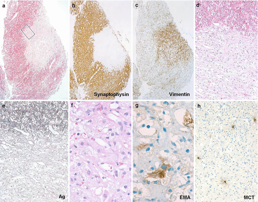
Hemangioblastoma is characterized by vascular proliferation of capillaries of variable size, along with a variable degree of stromal cells containing pink to clear foamy and lipid-laden cytoplasm (Fig. 32.2). Cystic change is commonly observed, yet necrosis is uncommon.
Histologically, hemangioblastoma may be difficult to differentiate from renal cell carcinoma or meningioma. Renal cell carcinoma and meningioma are typically immunopositive for EMA, however, whereas hemangioblastoma is not.
More specific and useful markers include podoplanin (D2-40) and inhibin A that are positive in hemangioblastomas but negative in renal cell carcinomas.

Fig. 32.2


Well-circumscribed hemangioblastoma (HBL) nodule partly surrounded by a crescent-shaped mantle of peritumoral pituitary parenchyma. (a) Optical contrast between the faint eosinophilic hue of the HBL nidus and the bright red granular quality of adjacent somatotrophs. (b, c) Adjacent section planes treated with immunohistochemistry, showing segregation of adenohypophyseal neuroendocrine cells (b) and mesenchymal-like immunophenotype (c) of the HBL nodule. (d) Detail view of boxed area in panel (a) shows the HBL comprise an irregular, reticular meshwork of tortuous, thin-walled capillaries that tend to be interspersed with pale stromal cells. (e) Gomori’s reticulin stain highlighting the brisk transition from the acinar outline of native adenohypophyseal follicles (upper third) to the vascular-dominated basement membrane pattern of HBL. (f) High-power view of HBL showing polygonal contours and cytoplasmic vacuolation of stromal cells encased by capillaries. Some nuclear pleomorphism, as also evident in this microscopic field, is of no prognostic significance. (g) A minority of stromal cells were stained for EMA. (h) Scattered mast cells are a characteristic complement of HBL. If not labeled otherwise, microphotographs have been made using H&E stain. Original magnifications: a–c, ×30; d, e, and h, ×100; f and g, ×400 (Adapted from Schar et al. [9]; with permission)
Stay updated, free articles. Join our Telegram channel

Full access? Get Clinical Tree


