Fig. 29.1
Hemangiopericytoma. (a) Sagittal T1-weighted gadolinium-enhanced image. (b) Coronal T1-weighted gadolinium-enhanced image. An enhancing mass is seen in the sellar and suprasellar region, arising from the region of the diaphragma sellae, with compression of the optic apparatus. The pituitary gland cannot be distinguished from the mass (Adapted with permission from Jalali R et al., Acta Neurochir (Wien) (2008) 150:67–71)
29.3 Histopathology
HPC is a mesenchymal, non-meningothelial tumor, closed related to the solitary fibrous tumor of the soft tissues [6, 7].
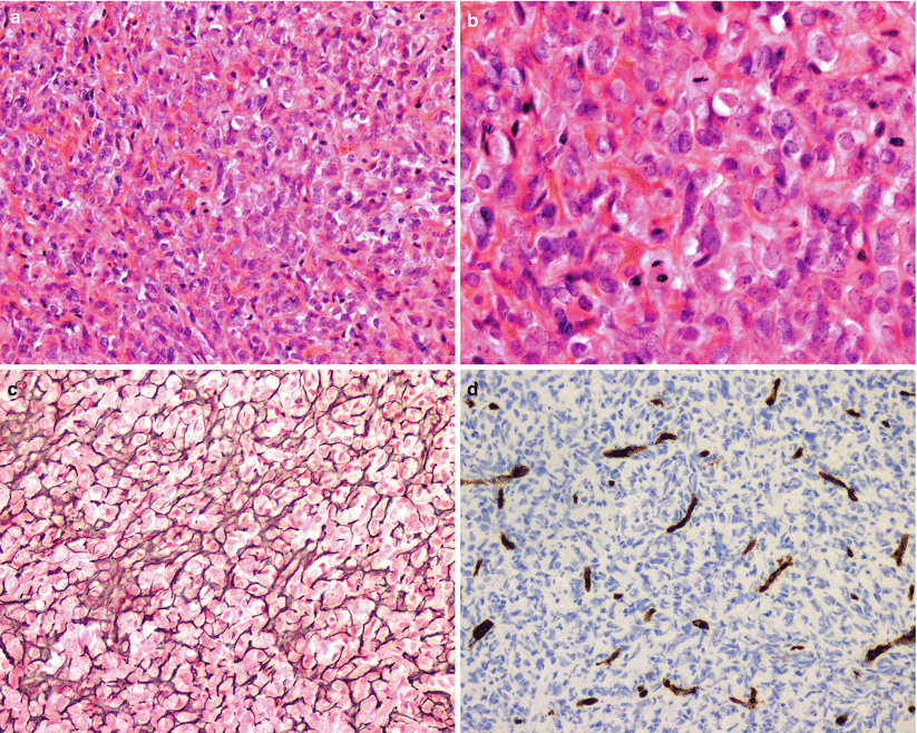
HPC is histologically characterized by high cellularity, closed apposed cells with round to ovoid nuclei arranged in a haphazard pattern, and intermixed by a rich network of thin-walled branching vessels (Fig. 29.2). Limited fibrous stroma is present.
In rare cases, malignant HPC can arise in the sellar region [8].
Sinonasal HPC is a distinct entity characterized by a diffuse, subepithelial proliferation of bland, uniform, closely packed spindle cells. A unique vascular network composed of variably sized, staghorn-appearing vascular channels, often with perivascular hyalinization, is often observed. Most cases are immunopositive for vimentin and smooth muscle actin staining [3, 9].

Fig. 29.2
Hemangiopericytoma. Hemangiopericytomas are hypercellular neoplasms composed of sheets of oval to spindle-shaped tumor cells with well-delineated cytoplasmic borders and oval and elongated nuclei (a, b). Mitotic activity may be obvious (b). A prominent network of delicate vascular clefts is seen (a), which is accentuated by immunoreactivity for reticulin (c) or endothelial cell immunomarkers like CD31 (d)
29.4 Clinical and Surgical Management
The primary treatment of skull base HPC is surgical resection, followed by radiation-based treatment for any residual disease. Endoscopic endonasal resection is the preferred approach for most sinonasal and sellar region HPCs [10].
HPC is an extremely vascular tumor. A priori knowledge of the diagnosis may offer an opportunity for preoperative embolization in selected cases.
Recurrence is noted in up to 92 % of patients following surgical excision. Complete excision is recommended whenever possible and is known to correlate with a lower recurrence rate [3, 11, 12].
Metastatic HPC has been reported in up to 20 % of cases.
Stay updated, free articles. Join our Telegram channel

Full access? Get Clinical Tree


