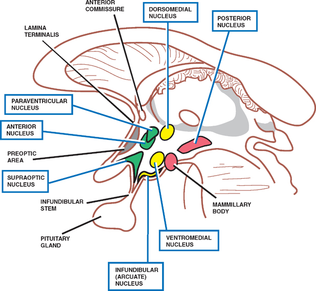
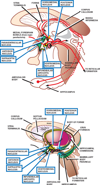
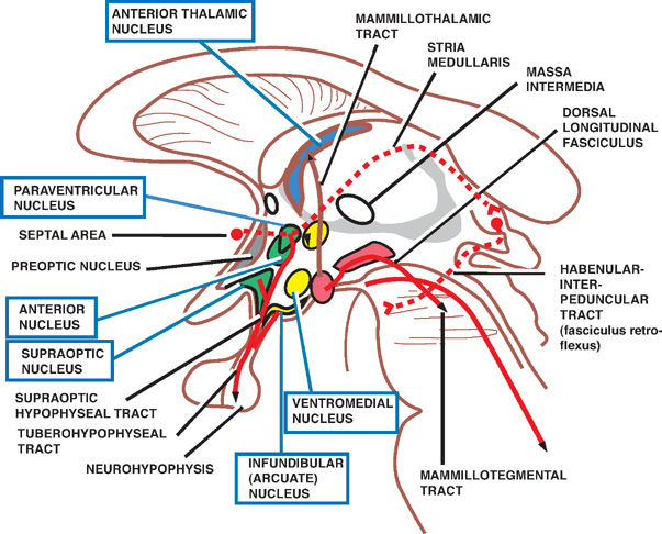
Hypothalamus

10 Hypothalamus


Hypothalamus


The hypothalamus is a small but crucial part of the dien-cephalon that lies below the thalamus and surrounds the lower part of the third ventricle.


From ventral to dorsal, the undersurface of the hypothalamus is marked by the optic chiasm, the tuber cinereum, the infundibulum, and the mammillary bodies.


The rostral border comprises the anterior commissure and the lamina terminalis; the caudal border merges with the tegmentum of the midbrain.


Laterally, the hypothalamus is flanked by the internal capsule; medially, it is flanked by the third ventricle.


The hypothalamus exerts its influence through three major systems: (1) the limbic, (2) the autonomic, and (3) the endocrine. It is composed of numerous nuclei of ill-defined boundaries that make connections with many parts of the central nervous system (CNS), including (1) limbic structures, (2) autonomic nuclei of the brainstem and spinal cord, and (3) the pituitary gland.


Although specific functions have not yet been assigned to individual hypothalamic nuclei, several afferent and efferent pathways of the hypothalamus have been described, and many functions have been identified.


This chapter describes the hypothalamic nuclei; the afferent and efferent connections of the hypothalamus; the limbic, autonomic, and endocrine hypothalamic functions; and the clinical manifestations of selected hy-pothalamic disorders.


Hypothalamic Nuclei


See Fig. 10.1.


The hypothalamus may be divided into a medial hy-pothalamic region that contains the majority of nuclei and a lateral hypothalamic region that contains the major fiber tracts (e.g., the medial forebrain bundle) and a group of diffuse nuclei.


The medial hypothalamic area is further subdivided into three regions: (1) the supraoptic region, which lies farthest anterior and includes the supraoptic, suprachias-matic, and paraventricular nuclei; (2) the tuberal region, which lies just posterior to the supraoptic region and includes the ventromedial, dorsomedial, and infundibular nuclei; and (3) the mammillary region, which lies farthest posterior and includes the mammillary body to the posterior nucleus.


image

Fig. 10.1 Hypothalamic nuclei.


Afferent Connections


See Fig. 10.2.


The hypothalamus is located in the center of the lim-bic system and receives numerous projections from lim-bic system structures. In addition, the hypothalamus receives ascending afferents from the brainstem reticular formation and descending afferents from the thalamus and cerebral cortex.


The major afferent connections of the hypothalamus include the following:


  • Olfactory and septal areas These areas are concerned with smell and basic emotional drives. They send axons to the hypothalamus via the medial forebrain bundle.
  • Hippocampus This limbic system structure is probably involved in a variety of behaviors, including learning and memory. The hippocampus sends axons to the mammillary bodies of the hypothalamus in a large fiber bundle called the fornix.
  • Amygdaloid nucleus Like the hippocampus, the amygdaloid nucleus is associated with complex behaviors. It sends axons to the hypothalamus via the stria terminalis.
  • Midbrain tegmentum (reticular formation) This includes a diffuse network of neurons concerned with a variety of autonomic functions. It sends axons to the hypothalamus through the medial forebrain bundle. Two additional brainstem nuclei, closely related to the reticular formation, also send axons to the hypothalamus. They are the raphe nucleus, which projects serotonin-containing fibers, and the nucleus ceruleus, which projects norepinephrine-containing fibers. Both fiber types project to the hypothalamus in the dorsal longitudinal fasciculus.
  • Dorsomedial and midline thalamic nuclei These are concerned with emotional states and autonomic functions. They project axons to the hypothalamus via the thalamohypothalamic tract.

image

Fig. 10.2 Afferent hypothalamic connections.


Efferent Connections


See Figs. 10.3 and 10.4.


The hypothalamus is a major output pathway of the limbic system. The efferent connections of the hypothalamus are largely reciprocal to the afferent projections. There are ascending connections to the cortex and thalamus as well as descending connections to the autonomic nuclei of the brainstem and spinal cord. Separately, two hypothalamic pathways project to the pituitary gland.


The major efferent connections of the hypothalamus include the following:


  • Olfactory and septal areas These areas are concerned with smell and basic emotional drives. They receive hypothalamic axons via the medial forebrain bundle.
  • Anterior thalamic nucleus This nucleus is the tha-lamic part of the limbic system. It is concerned with emotional states and memory. It receives hypotha-lamic axons via the mammillothalamic tract and projects in turn to the cingulate gyrus.
  • Preganglionic autonomic neurons of the brainstem and spinal cord These are concerned with various autonomic functions. They include the dorsal nucleus of the vagus (brainstem) and the intermediolateral cell column (spinal cord). Both receive input from the hypothalamus via the dorsal longitudinal fasciculus; the hypothalmic projections to the intermediolateral cell column are relayed in reticulospinal pathways.
  • Posterior pituitary gland The posterior pituitary gland receives direct axonal projections from large neurosecretory cells in the paraventricular and su-praoptic nuclei of the hypothalamus. They are carried in the supraoptic hypophyseal tract. Neurons of the paraventricular and supraoptic nuclei synthesize and secrete the hormones oxytocin and antidiuretic hormone (ADH), which are involved in reproductive functions and water balance, respectively. They are discussed in greater detail later in the chapter.
  • Anterior pituitary gland Neurosecretory cells in the infundibular nucleus of the hypothalamus produce releasing and inhibiting factors that influence the secretion of pituitary hormones. There are no direct axonal connections between the hypothalamus and the anterior pituitary gland. These factors are carried by axoplasmic transport in the tuberoinfundibular tract and are secreted into a capillary bed in the median eminence. From the median eminence, they are transported in hypophyseal portal veins to a second capillary bed in the anterior pituitary gland, where they modulate the secretion of trophic hormones, such as thyroid-stimulating hormone (TSH), adreno-corticotropic hormone (ACTH), follicle-stimulating hormone (FSH), luteinizing hormone (LH), growth hormone (GH), melanocyte-stimulating hormone (MSH), and prolactin.

image

Only gold members can continue reading. Log In or Register to continue

Stay updated, free articles. Join our Telegram channel

Dec 16, 2016 | Posted by in NEUROLOGY | Comments Off on Hypothalamus

Full access? Get Clinical Tree

Get Clinical Tree app for offline access