59 Interspinous Process Decompression
I. Key Points
– Lumbar stenosis refers to compression of the thecal sac, which may result in lower extremity pain/numbness (i.e., neurogenic claudication).
– Interspinous devices (ISDs) are designed to perform an “indirect” decompression by maintaining flexion of a stenotic segment, which increases the dimensions of the spinal canal and foramina.
– Appropriate candidates for ISD should experience clear relief of their claudication with lumbar flexion (i.e., sitting).
– This technique may give rise to reduced surgical morbidity and more rapid rehabilitation compared with laminectomy with or without arthrodesis.
II. Indications
– Symptomatic neurogenic claudication secondary to spinal stenosis with or without spondylolisthesis (confirmed by computed tomography [CT] or magnetic resonance imaging [MRI]) that has failed to respond to conservative treatments (e.g., physical therapy, medications, or epidural injections)
– Limiting lumbar extension may also target various sources of low back pain by unloading the posterior disc and facet joints.1
– Contraindications
• Significant spinal deformities (spondylolisthesis >grade I, scoliosis >25 degrees)
• Bony ankylosis
• Severe osteoporosis
• Critical stenosis/cauda equina syndrome
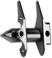
– The X-Stop spacer (Medtronic, Memphis, TN) is FDA-approved for use at one or two levels of the lumbar spine (Fig. 59.1).
III. Technique
– Preoperative planning
• Radiographs: evaluate for presence of spinal deformity (scoliosis on anteroposterior (AP), spondylolisthesis on lateral) and anklyosis (no segmental motion on flexion/extension views).
• CT/MRI: confirm diagnosis of stenosis.
• Dual x-ray absorptiometry (DEXA): assess risk for osteoporotic fractures.
– Anesthesia: general, monitored anesthesia care (MAC), or local
– Positioning: lateral decubitus or supine on a radiolucent table; make sure that the lumbar spine is maintained in flexion
– A vertical midline incision centered over the affected segment(s) is made through the skin and fascia, with care taken to avoid attenuating the supraspinous/interspinous ligaments.
– After the levels are confirmed using fluoroscopy or intraoperative x-rays, a subperiosteal exposure of the spinous processes and laminae is performed without disrupting the facet capsules.
– The interspinous space is distracted so that the ISD may be inserted between two adjacent spinous processes (depending on the surgical protocol for each specific implant).
– May also be combined with a microdecompression (e.g., laminotomies)
– The wound is closed in layers, and a drain may or may not be used.

Fig. 59.1 Photograph of X-Stop device, Medtronic Spine, LCC, Memphis, TN. (© Medtronic Spine LLC).
IV. Complications
– Intraoperative
• Spinous process (SP) fracture
• Malpositioning of implant
• Inability to safely place the implant (e.g., attenuation of ligaments, excessively small interspinous space secondary to “kissing” SP or facet hypertrophy)
– Postoperative
• Device migration/dislocation
• Persistent/recurrent symptoms (same-versus adjacent-segment degeneration)
– In the series of Barbagallo et al, the incidence of complications was 10.1% (primarily spinous process fractures and device dislocations) with a reoperation rate of 7.2%.2
V. Postoperative Care
– May be performed in ambulatory setting as opposed to inpatient admission
– Immediate postoperative ambulation
– No bracing typically required
– Gradual return to normal activities
VI. Outcomes
– Kuchta et al published the 2-year results of 175 consecutive X-Stop procedures performed at a single center.3
• X-Stop brought significant improvement in clinical outcome measures.
• Reoperation rate of 4.6% (removal of implant with posterior decompression)
– Zucherman et al reported the results of a multicenter, prospective, randomized trial comparing X-Stop and nonoperative modalities for spinal stenosis (191 patients).4
• At 2 years, clinical outcomes of patients receiving X-Stop were significantly improved compared with those undergoing conservative treatments.
• No device-related complications, but 6% of X-Stop cohort required subsequent laminectomy
– There continues to be a paucity of data on other ISDs.
VII. Surgical Pearls
– Appropriate candidates for this technique should experience clear relief of their claudicatory symptoms with lumbar flexion or sitting.
– Flexion-extension lateral x-rays may identify the presence of bony ankylosis at the stenotic level(s), which may preclude the implantation of an ISD.
– The lumbar spine should be maintained in flexion to facilitate intraoperative distraction of the stenotic segments
– Hypertrophic facet joints may need to be partially excised to allow successful placement of the ISD.
– Care must be taken when inserting the ISD in osteoporotic patients, who may be at greater risk for spinous process fractures.
Common Clinical Questions
1. Which of the following patients is best suited for placement of an ISD?
A. Patient with severe low back pain and minimal extremity symptoms
B. Patient with claudication secondary to moderate stenosis and a grade II spondylolisthesis at L4-L5
C. Patient with moderate stenosis at L3-L4 whose symptoms improve with sitting
D. Patient with severe stenosis between L2 and L5 who complains of perianal numbness and bladder dysfunction
2. What is the primary mechanism by which ISD relieves claudicatory symptoms?
A. Decreases facet joint forces
B. Unloads posterior disc tissue
C. Eliminates redundant ligamentum flavum and other compressive lesions
D. Increases the dimensions of the spinal canal
3. Which is not a typical characteristic of neurogenic claudication?
A. Pain is worse when walking uphill
B. Symptoms improve with sitting
C. Pain/numbness is worse with lumbar extension
Stay updated, free articles. Join our Telegram channel

Full access? Get Clinical Tree


