Intracranial Pressure and Neurocritical Care Monitoring
Charles L. Francoeur
Stephan A. Mayer
INTRODUCTION
Monitoring plays a fundamental role in the daily practice of neurocritical care. Bedside neuromonitoring supplements clinical evaluation and imaging with the underlying goal of detecting physiologic derangements before neurologic deterioration and irreversible damage occurs. This chapter introduces continuous bedside neuromonitoring modalities that are in use in the most advanced neurocritical care units around the world. Although no study has tested whether “multimodality” monitoring improves outcome, these advanced systems allow for recognition of real-time pathologic events that we were unable to identify just a few years ago and provide new insights into the complex pathophysiology of severe brain injury.
INTRACRANIAL PRESSURE MONITORING
PHYSIOLOGIC PRINCIPLES
Monro-Kellie Doctrine and Intracranial Pressure
The rigid skull is filled with incompressible content, namely brain (1,400 mL), cerebrospinal fluid (CSF) (150 mL), and blood (75 mL). In normal state, with blood outflow being equal to blood inflow, CSF absorption equals CSF production (˜20 mL/h) and intracranial pressure (ICP) lies between 5 and 15 mm Hg in the supine position (8 to 20 cm H2O).
If any one of those compartments increases in volume, as in the case of obstructive hydrocephalus, or a new mass lesion is added (a subdural hematoma for example), buffering mechanisms are called upon. The first adaptive process is egress of CSF in the spinal canal, provided there is no obstruction to flow. The second step is shifting of blood from the capacitance vessels (veins) out of the intracranial compartment. Once these buffering mechanisms are exhausted, intracranial compliance is reduced and ICP quickly rises (Fig. 33.1).
Secondary neurologic injury occurs with ICP over 20 mm Hg, and elevated ICP above this threshold after traumatic brain injury (TBI) or subarachnoid hemorrhage (SAH) correlates with increased mortality. The magnitude of ICP elevation (especially >40 mm Hg), the duration of intracranial hypertension, as well as its refractoriness to treatment are all associated with higher mortality.
Cerebral Perfusion Pressure
Arterial blood carries oxygen and glucose, which are necessary for neuronal function and survival. Cerebral perfusion pressure (CPP), defined as the difference between mean arterial pressure and ICP, is the main determinant of cerebral blood flow (CBF):
CPP = MAP – ICP
CPP values below 50 to 60 mm Hg can begin to cause cerebral ischemia, and systemic hypotension strongly correlates with mortality in TBI patients.
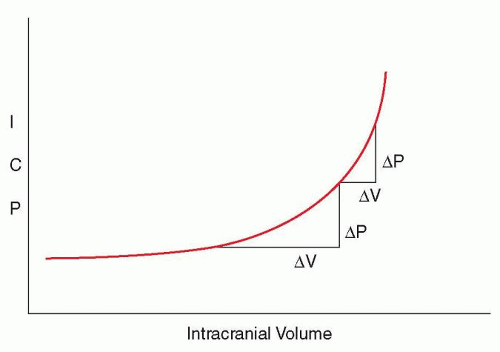
FIGURE 33.1 Compliance is the change in pressure per change in unit of volume (ΔP/ΔV). Once compensatory mechanisms are overwhelmed, compliance dramatically decreases, meaning that with a smaller increment in intracranial volume, a much more dramatic increase in pressure develops.
Intracranial Pressure Waveform
ICP is pulsatile with pressure deflections that correspond to transient increases in cerebral blood volume with systole. Bedside displays will show an ICP waveform with three components (Fig. 33.2): an initial percussion wave which occurs at the start of systole and under normal conditions in greatest amplitude (called P1), a secondary tidal wave then occurs which reflects brain recoil or elastance (P2), followed by a third tidal wave created by closure of the aortic valve at the start of diastole (P3). In states of reduced intracranial compliance, the ICP pulse pressure typically increases, and the amplitude of P2 exceeds that of P1 in the ICP waveform, as the “shock” of systolic inflow becomes less absorbable.
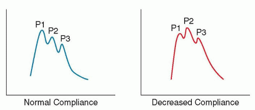
FIGURE 33.2 ICP waveform. P1, the percussive wave, comes from arterial transmission through the choroid plexus. P2, the tidal wave, reflects brain tissue elastance. As it rises, so does P2. When P2 exceeds P1, it has an excellent sensitivity to predict incoming increase in ICP. The third and final wave (P3) is secondary to aortic valve closure, a corollary of the arterial dicrotic notch.
Pathologic Intracranial Pressure Elevations
In patients with raised ICP, pathologic ICP waveforms may occur. Lundberg A waves (or plateau waves) represent prolonged periods (> 10 minutes) of high ICP (>20 mm Hg) (Fig. 33.3). They are caused by sustained vasodilation and abruptly occur when either CPP or intracranial compliance are low (see also Figure 107.4). Severe plateau waves preceding the onset of brain death can last for hours and reach levels as high as 50 to 100 mm Hg. Lundberg B waves are shorter duration (<10 minutes), lowamplitude elevations (<20 mm Hg) that indicate that intracranial compliance is compromised.
EXTERNAL VENTRICULAR DRAINAGE
Since the landmark study published by Lundberg in 1960, external ventricular drainage (EVD) has been the gold standard for ICP monitoring. It consists of a catheter blindly inserted into the lateral ventricle and connected via fluid-filled tubing to an external pressure transducer. Insertion is usually on the right side just anterior to the coronal suture roughly in the midpupillary line approximately 6 cm deep, although other approaches are described. The fluidfilled system relays the pulsatile waveform to a transducer, which is then converted to a digital signal. The ICP number displayed at the bedside represents the mean pressure. The hydrostatic reference point is the Foramen of Monro, which is estimated using the external acoustic meatus or the tragus.
EVD placement has the advantage of allowing therapeutic CSF drainage, making it the monitoring of choice in case of elevated ICP secondary to hydrocephalus. Depending on the indication, height of the EVD drip chamber is kept from 0 to 20 cm above the tragus, with lower levels causing a higher hourly CSF drainage rate. With a conventional EVD system, either ICP can be monitored with the system clamped or CSF can be drained but not both at the same time. Some EVD systems are equipped with a fiberoptic pressure transducer at the tip, which allows for simultaneous pressure monitoring and CSF drainage.
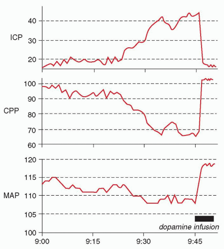
FIGURE 33.3 Lundberg A (plateau) wave, with characteristic “mirror” reduction in CPP. The plateau wave is terminated by an infusion of dopamine, which leads to an elevation in mean arterial pressure and reversal of the brain’s vasodilated state. ICP, intracranial pressure; CPP, cerebral perfusion pressure; MAP, mean arterial pressure.
The main complications of EVD placement are insertional bleeding and infection. Clinically significant (requiring intervention) bleeding rates are 1% or less, but the risk of infection (ventriculitis) ranges from 5% to 15%. As for any other invasive devices, aseptic insertion technique, limited manipulations, and quick removal are the best ways to keep infectious complications to a minimum.
Safe discontinuation of an EVD is accomplished by performing a 24-hour clamp trial. If the ICP remains below 20 mm Hg, the patient remains stable, and a follow-up CT shows no interval ventricular enlargement, the EVD can be safely removed.
INTRAPARENCHYMAL INTRACRANIAL PRESSURE MONITORS
Intraparenchymal devices are now the most commonly used method of ICP monitoring, and they are as reliable and accurate as EVD. In most systems, a fiberoptic or strain gauge pressure transducer is located at the catheter tip, which is inserted 0.5 to 1 cm into the cerebral parenchyma. Although often placed on the nondominant side, insertion should be ipsilateral to the lesion aiming for the brain parenchyma most at risk of secondary injury. The risk of insertional bleeding with a parenchymal ICP monitor is lower than that of an EVD, and the risk of infection is negligible.
INDICATIONS FOR INTRACRANIAL PRESSURE MONITORING
As a general rule, there are here indications for placement of an EVD or ICP monitor:
The patient is comatose (generally Glasgow Coma Scale [GCS] score ≤8).
Brain imaging indicates that the patient is at risk for elevated ICP due to the presence of significant intracranial mass effect.
Aggressive intensive care unit (ICU) care is warranted.
The Brain Trauma Foundation issued updated recommendations in 2015 regarding indications for ICP monitoring of severe TBI patients (Table 33.1). Other considerations include when clinical exam is likely to be lost for a moderate to long period of time, especially if associated with physiologic derangement, for example, during major surgery or a difficult to ventilate acute respiratory distress syndrome (ARDS) patient. Obstructive hydrocephalus is also a clear indication for EVD insertion. Recent guidelines from the International Multidisciplinary Consensus Conference on Multimodality issued recommendations for ICP monitoring in non-TBI patients (Table 33.2). Examples given in the consensus include massive ischemic stroke, meningitis, hypoxic-ischemic injury, and hepatic fulminant failure. Any pathology complicated by elevated ICP might benefit of such monitoring when clinical exam is not trustworthy, as it allows optimization of cerebral hemodynamics through CPP manipulation, aggressive ICP treatment, as well as detection of new catastrophic events.
TABLE 33.1 Indications for Intracranial Pressure Monitoring in Traumatic Brain Injury Patients
Level of Evidence
Indication
Level I and II A
There is insufficient evidence to support a level I or II A recommendation for this topic.
Level II B
Management of severe TBI patients using information from ICP monitoring is recommended to reduce in-hospital and two-week post-injury mortality.
From Guidelines for the Management of Severe Traumatic Brain Injury, 4th Edition; Brain Trauma Foundation, 2015.
EFFECT OF INTRACRANIAL PRESSURE-DIRECTED THERAPY ON OUTCOME
There is a strong association between elevated ICP, especially if refractory to treatment, and poor outcome. Retrospectives studies, databases analysis, and a meta-analysis show that 20 mm Hg is a powerful threshold for identifying patients at risk for death or poor outcome after TBI and also suggest a mortality benefit with ICP monitor-based management.
Of course, a monitoring device alone cannot improve the outcome of a disease. Only a treatment protocol based on the results of monitoring can impact on survival and recovery.
In 2012, the Benchmark Evidence from South American Trials: Treatment of Intracranial Pressure (BEST TRIP) trial compared two ICP treatment protocols, one triggered by ICP values exceeding 20 mm Hg and the other triggered by clinical examination and serial imaging results alone [Level 1].1 Outcome was similar with a tendency toward more efficiency in the ICP monitor arm (less hypertonic saline, less hyperventilation, less barbiturates). The external validity was problematic with poor prehospital care and even worse postdischarge management with few to no patients receiving rehabilitation. The medical community still believes ICP monitoring is an essential component of severe TBI management, although the possibility has been raised that perhaps more sophisticated triggers of therapy that take into account the state of autoregulation and intracranial compliance need to be explored. Current management algorithms for treatment of ICP are presented in Chapter 107.
TABLE 33.2 Indications for Intracranial Pressure Monitoring in Nontraumatic Brain Injury Patients
Level of Evidence
Indication
Moderate
Patients at high risk or those with clinical or radiologic evidence of acute symptomatic hydrocephalus
Low
SAH, ICH, and other non-TBI conditions in patients at risk for elevated ICP based on clinical and/or imaging features
Low
All poor-grade SAH patients with consideration for multimodality monitoring
Low
Patients who undergo hemicraniectomy in the setting of cerebral edema