Fig. 49.1
Juvenile nasopharyngeal angiofibroma (JNA). (a) Sagittal contrast-enhanced CT image. (b) Axial contrast-enhanced CT image. (c) Coronal contrast-enhanced CT image. (d) Sagittal precontrast T1-weighted MR image. (e) Axial T1-weighted post-gadolinium, fat-saturated MR image. There is an avidly enhancing mass in the posterior right nasal cavity and pterygopalatine fossa, eroding the floor of the right middle cranial fossa (c) and the clivus (d) and extending into the sphenoid sinus. (f) Digital subtraction cerebral angiography with the catheter in the right internal maxillary artery, showing markedly increased vascularity within the tumor
49.3 Histopathology
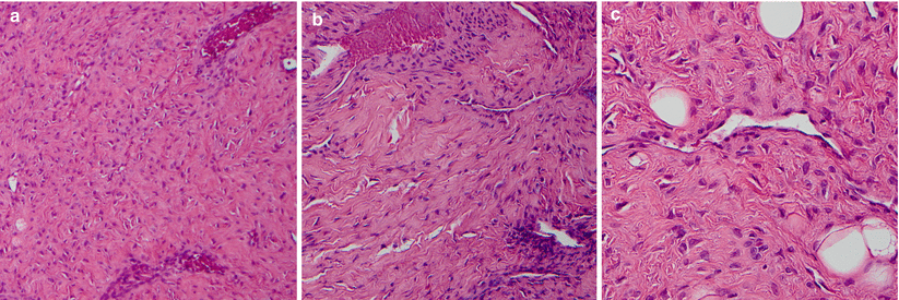
JNA is characterized by highly vascular capillary-like channels within a fibrous stroma (Fig. 49.2). Blood vessels typically lack smooth muscle and elastic fibers. Staghorn-shaped vessels may be seen.
Immunostaining for vimentin and vascular endothelial growth factor (VEGF) is typically positive [9].
Immunostaining for c-kit is almost always negative [9].
Immunoreactivity for the testosterone and estrogen receptors may play a key role in development.

Fig. 49.2
JNA. (a) Low-power view (×10 objective) of an angiofibroma, showing haphazardly arranged fibrous tissue and thin, wavy collagen. (b) Medium-power (×20) view of the same lesion showing multiple thin-walled blood vessels compressed by fibrous tissue. (c) High-power (×40) image of thin-walled blood vessel with a flattened endothelial cell lining surrounded by fibrous tissue containing cells with thin strands of wavy collagen. This lesion also had associated adipose tissue, evidenced by the presence of several mature adipocytes, and could be seen infiltrating adjacent adipose tissue (not shown)
49.4 Clinical and Surgical Management
The primary treatment for JNA is surgical resection. Possible approaches include open craniofacial, orbitozygomatic, endoscopic-assisted, and pure endonasal endoscopic approaches. In recent years, endoscopic approaches have been more widely explored for this indication [1, 7, 10, 11].
JNAs are often tumors highly vascular, firm, and rubbery.
Preoperative embolization of JNAs is an effective strategy for reducing intraoperative blood loss associated with resection [3, 12].
Neoadjuvant use of the testosterone receptor blocker flutamide has demonstrated some success in the preoperative treatment of JNA [13].
Although some degree of selection bias likely exists regarding assignment of the approach to varying tumors, endoscopic approaches have been associated with decreased intraoperative blood loss and lower recurrence rates [10].
Complete resections may be possible in over 90 % of patients; they are generally limited by the degree of internal carotid artery involvement. For higher-stage JNAs, neoadjuvant treatment or primary radiation may be a safe alternative [7].
Radiation therapy may be effectively employed for residual or recurrent disease and typically offers reductions in tumor volume of 30–50 %. Local tumor control is achieved in up to 85 % of patients [14–17].
Most patients have excellent outcomes, with 86 % experiencing disease freedom at a mean follow-up time of 47 months [12].
Stay updated, free articles. Join our Telegram channel

Full access? Get Clinical Tree


