60 Lumbar Arthroplasty
I. Key Points
– Lumbar arthroplasty has recently been approved in the United States as an alternative treatment strategy to spinal arthrodesis in the treatment of refractory symptomatic discogenic back pain.
– Etiology of low back pain remains unclear, with numerous possible pain generators.
– Goals: preserve physiologic spinal motion, prevent facet arthropathy, prevent adjacent-segment disease, restore disc height, and provide long-term pain relief1
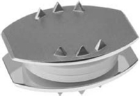
– The prosthesis should approximate the size and motion of the physiologic disc, avoid distracting the facet joints, and, ideally, reproduce the normal biomechanics (Fig. 60.1).
– No independent long-term, randomized, prospective study on any artificial disc has been published to date that clearly delineates the safety and efficacy of lumbar arthroplasty.
II. Indications
– Patient refractory to a minimum of 6 months of conservative, nonoperative treatment
– One- or two-level symptomatic degenerative disc disease in patients of age 18 to 60 years
– Correlative objective radiographic findings including disc desiccation, vacuum disc, high-intensity zone signal, and Medic signal changes
– Postdiscectomy axial back pain or juxtafusion disc degeneration
– Absence of central or lateral recess stenosis that may require posterior decompression to address concomitant radicular leg pain
– Provocative discography may demonstrate concordant pain reproduction and confirm diagnosis.

Fig. 60.1 Photograph of the Charite artificial disc (with permission from DePuy Spine, Inc.).
III. Contraindications
– Compromised structural integrity of bone
• Tumor, osteoporosis (T-score <–2.5), osteomalacia, acute fracture
– Potential compromise of stability or alignment of implant
• Scoliosis, spondylolysis, spondylolisthesis (>grade I), incompetent posterior elements
– Conditions that may compromise clinical outcome of disc replacement
• Significant facet arthrosis, disc herniation with predominant radicular symptoms, central or lateral recess stenosis, disc height less than 5 mm
– Miscellaneous
• Obesity (BMI >40), local or systemic presence of tumor or infection, pregnancy, intraspinal neoplasm
IV. Technique2
– Positioning
• The patient is placed in the supine position on a radiolucent table with all bony prominences padded after institution of general anesthesia and Foley insertion to decompress the bladder.
• C-arm fluoroscopy is used to identify the approach angle and location of the disc space and to verify that clear anteroposterior and lateral images can be attained.
– Standard sterile preparation and draping are performed and prophylactic intravenous antibiotics are administered.
– Exposure
• The majority of prostheses are implanted by means of an open anterior approach similar to that used for anterior lumbar interbody fusion (ALIF).
• A general or vascular surgeon can obtain access to the spine through either an anterior retroperitoneal approach (most preferred) or a midline transperitoneal approach.
• A left-sided approach is preferred between L3 and L5 due to the relative resilience of the aorta and ease of mobility compared with the vena cava.
• In accessing L5-S1 in male patients a right-sided approach is recommended to reduce the rate of injury of the superior hypogastric plexus, which can cause retrograde ejaculation.
• The retroperitoneal space is entered deep to the rectus sheath and the peritoneum, with the ureter retraced medially.
• Blunt dissection reveals the lateral edge of the psoas, and vascular structures are carefully mobilized and elevated off the anterior spine.
– Procedure
• Fluoroscopic images are used to confirm the disc level and identify the midline.
• A subtotal discectomy is performed by excising the anterior longitudinal ligament, annulus, and nucleus pulposus, leaving the lateral portion of the annulus intact.
• The cartilaginous end plate is debrided from the osseous end plate. The integrity of the end plate is preserved to ensure implant fixation and avoid subsidence.
• If necessary, the posterior longitudinal ligament (PLL) is released and posterior osteophytes debrided.
• Special instruments are used to measure the footprint, lordotic angle, and core height.
• Based on preoperative templating and intraoperative sizing, the appropriate trial is inserted.
• The appropriately sized prosthesis is implanted after device-specific preparation of the end plate.
• If the device has a polyethylene core, it is trialed and inserted after confirming on lateral fluoroscopy the restoration of desired disc height and lordosis.
– Fluoroscopy confirms the central position of the implant on the anteroposterior view. Ideally, the center of rotation of the device is 2 mm posterior to the sagittal midline of the vertebral body on the lateral view.
– Final confirmatory radiographs are obtained and the wound is closed in routine fashion.
– The approach varies according to the lumbar level accessed as well as device-specific modifications to the general technique.
V. Complications
– Approach-related complications (10 to 13%): vascular injury, phlebitis, pulmonary embolism, sexual dysfunction, and retrograde ejaculation.
– Postoperative retroperitoneal scarring makes revision surgery more difficult.
– Failure of the prosthesis primarily involves facet joint degeneration, subsidence, device migration, and adjacent-level disease.
– Cases of vertebral body fractures have been reported.
– Heterotopic ossification has been reported in varying degrees in 1.4 to 15.2% of patients.
– A prospective, randomized, multicenter Food and Drug Administration (FDA)–regulated clinical trial reported on complications of 589 patients.3
• The disc replacement group was found to have an 8.8% reoperation rate, compared with 10.1% in the lumbar fusion control group. Mean time to reoperation was 9.7 months.
• The primary reason for removal of the implant was device migration (75%).
• A higher incidence of vascular injury occurred in the reoperation group (16.7%) compared with the primary group (3.4%).
• Fourteen patients required posterior instrumented fusion for persistent low back pain (2.4%).
VI. Postoperative Care
– Patients bear weight as tolerated and are mobilized on the first postoperative day.
– A brace is typically not needed.
– Standing radiographs are obtained as soon as feasible postoperatively to document the position of the implant in the weight-bearing position.
– A gentle low back and abdominal strengthening program is implemented starting with the first postoperative day.
– The patient is given postoperative restrictions including avoidance of substantial extension, bending, twisting, or heavy lifting for the first 6 weeks.
– Progressive unrestricted activity is allowed after 6 weeks.
VII. Outcomes
– Results of a prospective, randomized, multicenter FDA-regulated trial conducted to assess the safety and efficacy of the procedure were recently published.4
• The study reported on 160 patients who completed 5-year follow-up.
• Patients were randomized to total disc replacement (TDR) or anterior interbody fusion using Bagby and Kuslich (BAK) cages with iliac crest autograft.
• Overall success was 57.8% in the TDR group versus 51.2% in the BAK group.
• Oswestry Disability Index (ODI), Visual Analogue Scale (VAS) pain scores, patient satisfaction, and SF-36 questionnaires were similar across the two groups.
• The mean range at the index level was 6 degrees for the TDR patients and 1 degree for the BAK patients. Changes in disc height were similar for the two.
• The authors concluded there is no statistical difference in clinical outcomes between the groups.
• The TDR patients did reach a statistically higher rate of employment and lower rate of long-term disability compared with the BAK patients.
– The clinical efficacy, long-term durability, and potential complications need to be more clearly elucidated with the use of unbiased prospective randomized studies with long-term follow-up.
VIII. Surgical Pearls
– The lumbar spine is positioned in the neutral position to minimize tension on retroperioteneal vessels.
– Sympathetic and parasympathetic nerves must be carefully preserved to prevent erectile dysfunction and retrograde ejaculation in male patients.
– The integrity of end plates must be preserved to ensure good implant fixation and avoid subsidence.
– The implant must restore appropriate lordosis, have adequate end plate coverage, and avoid distraction of more than 3 mm to apply proper tension to the posterior ligaments.
Common Clinical Questions
1. All of the following are contraindications to total disc arthroplasty except:
A. The presence of significant lateral recess stenosis
B. Facet arthrosis
C. Degenerative spondylolisthesis
D. Smoking
E. Obesity (BMI >40)
2. Which of the following statements is true with regard to total disc arthroplasty?
A. Randomized prospective control trials have illustrated the efficacy of lumbar disc arthroplasty, establishing that it preserves normal biomechanics and reduces the incidence of adjacent-segment degeneration.
B. A right-sided approach to the L5/S1 disc space is recommended in male patients undergoing total disc arthroplasty.
C. The PLL is essential to the appropriate tensioning of the disc replacement and should not be excised.
D. Heterotopic ossification is not a confirmed potential complication of total disc arthroplasty.
E. A significant portion of the cranial and caudal end plate must be removed prior to placement of the implant for proper setting of the prosthesis.
Stay updated, free articles. Join our Telegram channel

Full access? Get Clinical Tree


