51 Lumbar Laminectomy
I. Key Points
– An adequate lumbar laminectomy affords good central decompression and lateral gutter decompression, and sets the stage for foraminotomies at the corresponding spinal levels.
– Removing more than one-third of the medial facet joints during a laminectomy could accelerate degeneration and cause instability, necessitating a subsequent fusion.
II. Indications
– Focal or multilevel lumbar central and lateral recess stenosis
III. Technique
– Place patient in the prone position.
• A Wilson frame is adequate if no instrumentation is planned; otherwise an OSI table is generally used.
– Localize with fluoroscopy.
– Make a midline incision at the appropriate level and perform a subperiosteal dissection to detach the erector spinae muscles from the lamina.
– Stop at the medial aspect of the facet joint. Do not disturb the facet joint capsule.
– Use a Horsley or Leksell rongeur to remove the spinous process(es).
– Use a drill with an AM-8 bit to complete the laminectomy; alternatively, use Kerrison and Leksell rongeurs.
• The facets are often hypertrophied, and the medial one-third can be drilled or removed with Kerrison rongeurs.
– Know your landmarks and be careful to avoid drilling the facet joint, pedicle, or pars.
– Remove the ligamentum flavum with Kerrison rongeurs to complete the decompression.
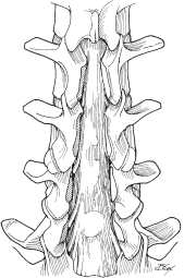
– Use a ball-ended probe to inspect the foramina (Fig. 51.1).
• Use the number 2 Kerrison if necessary to perform the foraminotomy.
– Use a subfascial drain if the wound is extensive.
– Ensure a tight fascial closure and close the skin in the standard fashion.

Fig. 51.1 Dorsal view of lumbar spine showing decompressed thecal sac and nerve roots with laminectomy taken to the medial facets laterally.
IV. Complications
– Cerebrospinal fluid leak (CSF) leak (0.3 to 13%; increases to about 18% in redo operations)1
• Should be repaired using nonabsorbable suture
– Neurologic deficit (0.3%)1
– Wound infection (1 to 5%)2
– Spondylolisthesis3
– Death (0.06%)1
V. Postoperative Care
– Mobilize early without brace.
– Remove drain when output drops below 50 ml per 8-hour shift (typically postoperative day 1).
– Discharge home when patient meets discharge criteria.
VI. Outcomes
– Review of literature reveals 60 to 70% with good to excellent results.1–4
– Reoperation in 10 to 30% for recurrent/adjacent-level stenosis, spondylolisthetic stenosis, or instability1,3–5
VII. Surgical Pearls
– With advanced degenerative disease, obtain preoperative dynamic films to rule out a dynamic spondylolisthesis.
– Leaving the ligamentum flavum until all the “bony work” is completed protects the dura during drilling and use of the Kerrison punches.
Common Clinical Questions
1. Why are preoperative dynamic x-rays sometimes obtained before a lumbar laminectomy?
2. During lumbar laminectomy, what location is associated with the highest likelihood of a CSF leak?
Stay updated, free articles. Join our Telegram channel

Full access? Get Clinical Tree


