Showing most revelant items. Click here or hit Enter for more.
Lumbar Puncture and Cerebrospinal Fluid Analysis
Nancy J. Edwards
Tareq Saad H. Almaghrabi
Kiwon Lee
INTRODUCTION
Accessing the cerebrospinal fluid (CSF) compartment—typically via lumbar puncture (LP)—is one of the key skills every neurologist should have. Proper analysis of CSF findings may result in a diagnosis (even if the neurologic examination and neuroimaging have not); furthermore, LP and the removal of CSF may be therapeutic in certain cases. This chapter will review step by step the technique of the LP, including advances in the procedure (e.g., ultrasound guidance); in addition, we outline here a guide for the interpretation of CSF findings in various neurologic disorders ranging from infectious to demyelinating to neoplastic.
LUMBAR PUNCTURE
TECHNIQUES
Careful positioning of the patient is often the key to performing a successful LP. Patients may be placed in either a lateral recumbent position with the spine flexed—head bowed, knees bent, and drawn up into the torso, for example, the “fetal position” (Fig. 31.1). Alternatively, patients may sit upright with the neck and back flexed forward (typically resting on a table). The lateral recumbent position should be used if an opening pressure is needed. As the conus medullaris ends at the level of the L1 and L2 interspace in 94% of adults, this level should be avoided during LP. Instead, the L3-L4, L4-L5, and L5-S1 interspaces are appropriate for needle puncture. These interspaces can be identified via palpation of bony landmarks. The line joining the superior aspect of the iliac crests posteriorly (the intercristal line) identifies the L4 spinous process or L3 and L4 interspace.
The interspace for needle insertion should be marked and the skin overlying the LP site should be sterilized and draped using standard aseptic technique. Local anesthetic—usually 1% lidocaine—is then applied; the 25-gauge needle should be used to raise a skin wheal at the LP site and the longer, 20-gauge needle should then be used to infiltrate the deeper layers. The spinal needle (with stylet in) is then inserted through the skin wheal into the interspinous space. Ideally, needle insertion should be midline, orthogonal to the plane of the back. To reduce the risk of post-LP headache, the bevel should be parallel to the longitudinal fibers of the dura; if the patient is in the lateral recumbent position, the bevel should face up and if the patient is sitting upright, the bevel should face to one side. Theoretically, this allows dural fibers to be separated rather than cut. The spinal needle is then advanced slowly at a slightly cephalad angle (directed toward the umbilicus) in order to follow the contour of the spinous processes. The needle will pass through the supraspinous ligament and the ligamentum flavum, perhaps resulting in a “pop” when the dura is pierced and the subarachnoid space is entered. The stylet should then be removed and CSF should readily appear at the needle hub if in the subarachnoid space. If instead CSF does not flow, the stylet should be replaced and the needle advanced or withdrawn incrementally, with frequent removal of the stylet, until CSF is obtained or bone encountered.
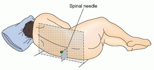
FIGURE 31.1 Proper positioning for lumbar puncture. Position the patient’s back at the edge of the bed with the head flexed and the legs curled up in the fetal position. Place a pillow under the head. Hips are parallel to each other and perpendicular to the bed. The spinal needle should be parallel to the bed. (Adapted from Marshall R, Mayer S. On Call Neurology. 3rd ed. Philadelphia: Saunders; 2007.)
ULTRASOUND GUIDANCE
Ultrasound visualization of landmarks may significantly improve the rate of LP success. This is particularly true in certain patient populations where palpation of landmarks may be challenging—neonates, pregnant women, obese patients, and patients with generalized edema, for example. One large meta-analysis of 14 studies and 1,334 patients revealed a significant reduction in the risk of failed or traumatic LPs and epidural catheterizations with the use of ultrasound guidance. The absolute risk reduction was 0.63, resulting in a number needed to diagnose of 16 to avoid one failed tap.
To perform an ultrasound-guided LP, the linear probe (higher resolution of superficial structures) should be used. The first view that should be obtained is the transverse view—the goal of this view is to determine an accurate anatomic midline by identification of the spinous process (Fig. 31.2A). To obtain this view, the probe is placed perpendicularly to the long axis of the spine. The spinous process will appear as a hyperechoic white convex rim with an anechoic shadow (or the anechoic shadow itself can simply be used if the rim is not identified). The midline should be marked and the longitudinal view should then be obtained (using the midline as a reference). The goal of the longitudinal view is to determine the spinal interspace—where the spinal needle will be inserted. To obtain this view, the transducer should be rotated into the sagittal/longitudinal plane with the probe marker pointing cephalad. Identify the hyperechoic spinous process again and then adjust the probe cephalad and caudad in order to center the probe/image between two contiguous, crescentic-shaped spinous processes; the interspace will be the hypoechoic gray gap in between (see Fig. 31.2B). This interspace should also be marked. Remove the probe and extend the transverse/longitudinal markings until they intersect; this intersection represents the ideal position for the spinal needle to be inserted.
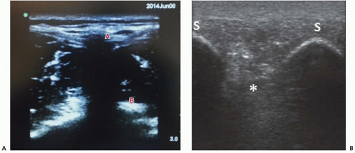
FIGURE 31.2 Ultrasound-guided LP. A: Transverse view. The anechoic shadow below A represents the spinous process—the midline; B localizes the transverse process. B: Longitudinal view. The interspace (asterisk) is located between the two crescentic-shaped, hyperechoic spinous processes (S).
CEREBROSPINAL FLUID ANALYSIS
CEREBROSPINAL FLUID PLEOCYTOSIS
Basic analysis of the CSF begins with red blood cell (RBC) and white blood cell (WBC) analysis and measurements of protein and glucose, although an extensive variety of additional tests can also be ordered (Table 31.1). The normal WBC count of CSF ranges from 0 to 5 lymphocytes or monocytes per cubic millimeter. Rarely, a single polymorphonuclear (PMN) cell may be identified from a large volume tap; the presence of two or greater PMNs, though, should be considered abnormal. A myriad of neurologic disorders can result in a CSF pleocytosis, ranging from acutely life-threatening (bacterial meningitis) to entirely chronic (as can be seen in patients with HIV).
Assessing the magnitude of pleocytosis along with the relative predominance of neutrophils to lymphocytes is often key, as is the clinical history—specifically whether the neurologic symptoms are acute or chronic in nature. For instance, a patient presenting with acute encephalopathy and a neutrophil-predominant CSF pleocytosis should be presumed to have bacterial meningitis (and promptly treated as such). In general, patients with acute bacterial meningitis have a CSF WBC count of several thousand (although the range may be from hundreds to >60,000). Furthermore, a CSF sample with greater than 33% neutrophils, a protein concentration of greater than 100 mg/dL, and a glucose level of less than 50% of the serum glucose level is strongly suggestive of bacterial meningitis. Exceptions include bacterial meningitis due to Listeria (often with a lymphocytic rather than neutrophilic pleocytosis), early viral or atypical (fungal/mycobacterial) infections that may initially recruit neutrophils into the CSF, neuroinvasive disease due to West Nile virus, and neuroinflammatory disorders (neuro-Behcet, Sweet syndrome).
In an acutely ill patient with a lymphocytic CSF pleocytosis, viral meningoencephalitis is often the cause. The WBC count generally ranges from 10 to 1,000 cells/mm3 and is composed primarily of mononuclear cells. The cornerstone of etiologic diagnosis is the detection of viral nucleic acid sequences in the CSF via polymerase chain reaction (PCR). In cases of suspected viral meningoencephalitis, CSF PCRs for the enteroviruses along with the herpesviruses (herpes simplex virus 1 and 2, cytomegalovirus, varicella-zoster virus, Epstein-Barr virus (EBV), and in immunocompromised patients, human herpesvirus-6) should be sent, along with serologic testing for HIV. Serologic testing for several additional viruses can be considered (West Nile virus, St. Louis encephalitis virus, Eastern equine encephalitis virus, Venezuelan equine encephalitis virus, and La Crosse virus).
TABLE 31.1 Common Cerebrospinal Fluid Tests
Cell count
Protein and glucose levels
Gram stain and culture
VDRL test
India ink test (for Cryptococcus neoformans)
Wet smear (for fungi and amebae)
Stain and culture for AFB (for tuberculosis)
Cryptococcal antigen titers
pH and lactate levels (abnormal in MELAS)
Oligoclonal bands (abnormal in multiple sclerosis)
IgG index (intrathecal IgG production)
IgG and IgM antibody titers (for viral infections, compare to serum)
Lyme disease antibody titers (compare with serum titers) and Western blot