Chapter 189 Management of Cauda Equina Tumors
Cauda equina tumors (CETs) are seldom reported and their specificity is seldom emphasized, and yet they ought to be considered as a distinct entity due to their anatomic localization, their surgical management, and their clinical course. Neurosurgeons should be aware of this surgical anatomic entity, and CETs need to be referred to well-trained teams. Some pathologies—lipomas, dermoid cysts and teratomas—are virtually specific to the pediatric population and are described in a separate section in this chapter.
Most CETs involve adolescents or young adults and are slow growing benign tumors arising from the nerve sheath. Some may be the expression of a congenital disease such as neurofibromatosis or von Hippel-Lindau disease. The diagnosis is established following a long time lapse if the initial symptoms are considered, leading to tumors of large or even giant size. This large size is also, like the previous feature, due to the wide free space surrounding the cauda equina. Postoperative functional status is correlated to the preoperative status of patients at the time of the operation.
Most of these tumors are benign, which justifies an aggressive surgical approach, using magnification techniques and microsurgical instrumentation. Adjuvant therapy is unnecessary, at least after the first procedure. In 1996 we performed a retrospective review of the French cases with 231 patients.1,2 Our personal series comprises 31 patients. It is not always easy to make a distinction between a true CET and a tumor arising from the neighboring structures, even with the magnetic resonance imaging (MRI) data, and it is sometimes necessary to obtain the operative and anatomopathologic findings to correctly classify the tumor. In the literature, many isolated cases are reported, but large series and practical advice for management are seldom encountered.
Definition of Cauda Equina Tumors
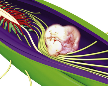
Primary tumors of the cauda equina arise from the different intrinsic structures of the region, such as the filum, the nerve sheaths, intrinsic vessels of the nerves, conjunctive tissue, and embryologic remnants.3 Some authors include tumors sprouting from the surrounding tissues—meningeal envelopes, epidural structures, and bone—but this does not seem to be the proper nosologic approach in description of this pathology, even if their clinical features present some similarities. It is in fact relatively difficult to classify metastatic tumors that, although not infrequent, do not constitute a surgical problem (Fig. 189-1).

FIGURE 189-1 Artist’s view illustrating the difficulty of classifying some cauda equina tumors depending on adjacent tissues and tumor connections.
(Graphic conception by J.P. Giot and P. Rigoard.)
In infants and children, CETs are often associated with occult dysraphism and are treated with their special features taken into account.
The most common tumors are benign schwannomas, neurofibromas (nerve sheath tumors), ependymomas in their myxopapillary type (filum tumor), hemangioblastomas, lipomas, epidermoid cysts, teratomas, and paragangliomas. Some others can develop on the nerve roots; these include lymphomas, metastases, and capillary hamartomas. Chordomas can develop inside the dura in the absence of any patent connection with the sacrum or the spine. Astrocytomas have also been reported, which develop inside the conus medullaris, involving the proximal part of the roots, but these are not true CETs.
Clinical Presentation
The involved population includes both sexes, with a slight male predominance, which is more marked for ependymomas (nearly 61%).1,2 Mean age for all CETs is 47 years, but there is a strong correlation between age and tumor type: 34.6 years ± 16 years for ependymomas and 51 years ± 17 years for benign neurinomas.
Symptoms at the time of diagnosis common to all tumors include low back pain (60%), radiculopathies (81%), and sphincter dysfunctions (25%). As a general rule, to the clinician should pay special attention to bilateral radicular pain. There are some differences linked to the pathologic type: Low back pain is seen in 70% of patients with ependymomas but only 55% of those with neurinomas. Radiculopathy is seen in 75% of patients with ependymomas and in 93% of those with neurinomas. Functional sphincter or genital disturbance is seen in 3% of patients with neurinomas and in 24% of those with ependymomas. Other symptoms are seldom associated with a specific type of tumor: focal deficit is seen in 33% and amyotrophy is seen in 8.5%, but these signs are never isolated.
Physical examination discloses a limited range of spinal motion in 50% of patients, a paravertebral spasm in 28%, abolition of one or several reflexes in half, and of this half, 25% have bilateral abolition of ankle tendon jerk and 13.5% have bilateral abolition of knee jerk. A motor deficit exists in 42% of cases, often bilateral at the L5 and S1 levels (15%) and less commonly at the L2, L3, and L4 levels. A sensory deficit exists in 50% of cases at the same levels. Amyotrophy was found in 15%. Saddle hypoesthesia or anesthesia exists in 20% of cases bilaterally and in 3.5% unilaterally, most often in patients with ependymomas. The bilaterality of symptoms mainly involves those with ependymoma. Associated genital and sphincter dysfunctions affect 34.2% of patients and needs to be precisely sought and explored, including urinary emergency, episodic incontinence, overflow with retention, and loss of anal and bulbocavernosus reflexes. All patients can walk, but some (11%) require a cane or a crutch.
The time interval between the first symptom and the diagnosis ranges from 1 month to 264 months. In our series the mean delay was about 34 months for 50% of the patients. There is no influence of age but rather a strong correlation with tumor type: long-lasting time lapse, 50 months for neurofibromas and paragangliomas; medium, 20 months for neurinomas and ependymomas; short for malignant tumors, especially metastasis. In a few cases intratumoral bleeding caused by a trauma results in an acute cauda equina syndrome.4
Diagnostic Evaluation
Electromyography and evoked potentials are of limited value in the investigation of CETs.
Plain X-Rays
Plain x-rays are taken in almost all patients, but they are unremarkable in two out of every three cases. They can reveal scoliosis, increased interpedicular distance, or scalloping in large lesions. This last sign is nonspecific and is only the mark of large chronic or slow-growing lesions; it is also encountered in large arachnoid cysts. Erosion of the posterior wall of a vertebral body can be observed in 21% of ependymomas and neurinomas.
Enhanced Computed Tomography Scan
Enhanced CT has been performed in 53.5% of our cases and was not contributive in half; this fact results from incomplete examination leading to a CT scan focused on the last three levels, below the level of the tumors. Sometimes misinterpretation arises when a spinal canal is completely filled by the hypodense tumor and the radiologist draws the conclusion that there is no compression. Contrast administration showed intraspinal enhancement in one case out of two and osseous abnomalities in one out of four. Water-soluble intrathecal injection allows enhanced visualization of lesions, including their size and shape.
Computed Tomography Myelography
Myelo-CT scan is used in patients for whom MRI is contraindicated or impossible to obtain. The whole lumbar spinal canal must be examined: Misdiagnosis was found in two cases of giant tumors with a CT scan limited to the three lowest vertebrae.
Magnetic Resonance Imaging
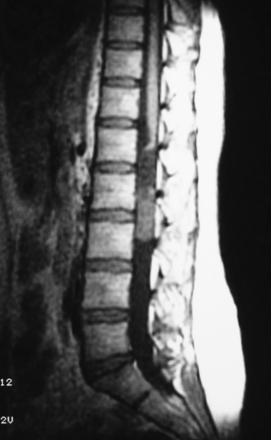
MRI is the gold standard for diagnosis. It must include T1- and T2-weighted sequences, without and with gadolinium administration, and in sagittal and axial views. It demonstrates the existence and the size of the tumor, cystic components, intratumor bleeding, vascularity, and the relation of the tumor to the conus, and it is contributive in 100% of cases. It also rules out other pathologies. It leads to a presumptive histopathologic diagnosis.5
Management Decisions
The diagnosis of a CET necessarily mandates operative removal following an exhaustive general and neurologic evaluation of the patient.
In young patients, if the diagnosis of neurinoma or neurofibroma is suspected, signs of neurofibromatosis must be sought. If several features of the disease are found, an extensive exploration of the central and peripheral nervous system is required.
The functional problems and prognosis have to be precisely described to the patient and his or her family, and the expectations must be realistic. Patients harboring minor neurologic impairment must be aware that neurologic deterioration can result from the operation, and in all cases hospitalization in a rehabilitation center will probably be necessary. The sphincter dysfunction is the least likely symptom to improve after surgery if it has existed for several weeks, and it may be amplified or appear postoperatively. As in intramedullary spinal tumors, the final outcome depends on the preoperative neurologic status of the patient. Because those tumors are nearly always benign, there is no alternative treatment to surgery. The goals of the operation are tumor removal and preservation or improvement of neurologic function.
Technical Adjuncts to Tumor Removal
The ultrasonic aspirator is less useful than in spinal cord tumors and may be dangerous, especially between S2 and S5, where the nerve roots are tightly applied to the tumor.
Laser may be used with low intensity, but classic microsurgery tools and optic magnification are usually sufficient.
Intraoperative stimulation of motor roots may be used for their identification when they are surrounded by tumor tissue.6 Evoked potential recordings are sometimes used, but they also are far less effective than in spinal cord tumors, and there is no evidence that they improve outcome.
Surgical Technique
Surgery of CETs requires 3 to 8 hours, and the patient is positioned prone on soft cushions, avoiding compression of the belly. A bladder catheter is always inserted, and infusion into a large vein. The head must be kept in flexion to avoid massive loss of CSF. The incision is median and must be 5 cm above and below the tumor limits.
Laminotomy must be performed systematically in infants and young adults, with reconstruction of a normal anatomy by reinserting the posterior processes prevents growth deformation in the young but also decreases the post-operative pain and the frequency of CSF leaks (Fig. 189-2).

FIGURE 189-2 Schematic drawings illustrating surgical approach via laminotomy. A, Lines of bone incision during laminotomy. The area of removal includes the spinous process and the laminae. B, Exposure obtained with intermyolaminar approach. 1, Spinous process; 2, projection of the tumor on the dura mater (dotted line); 3, the rostral and caudal flaps of the spinous process and laminae, which are lifted, will be put back in place and fixed; 4, laminae; 5, cutting line of the laminae; 6, incision of the dura mater. C, Opening of laminotomy after section of interspinous ligament. 1, Spinous process; 6, Supraspinous ligaments; 7, the opened lamina is fixed with wires.
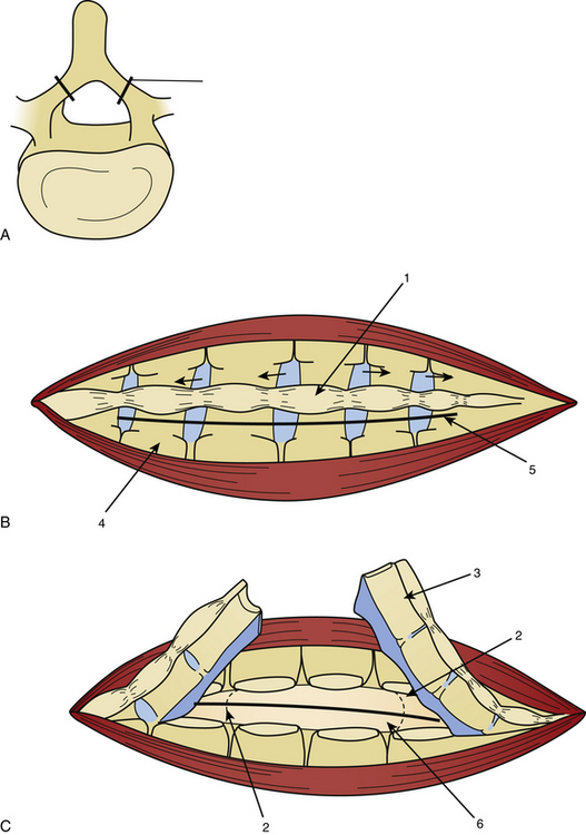
The dura mater may be absent in large ependymomas, and care must be taken not to injure the roots during the laminotomy. In the event of reoperation, laminectomy is unavoidable. The dura opening must allow control of the tumor’s extremities (Fig. 189-3), and the dura is fixed with stitches to the muscles. Dividing the tumor under optic magnification may begin. The best way to proceed is to start at the upper limit of the tumor, proceed inferiorly to the lower (Fig. 189-4), and to finish at the central part of the tumor. This strategy allows optimal root control (Fig. 189-5). Most of the roots are pushed against the dura by the tumor. Tumors of the filum terminale displace the roots on the right and left sides of the spinal canal, but they can develop between the roots, covering and hiding them. It is important to identify the filum, and stimulation can help do this.

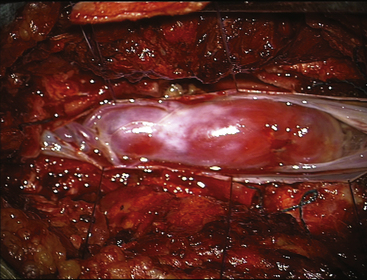
FIGURE 189-3 Operative view showing exposure of both extremities of a cauda equina paraganglioma after opening of the dura mater.

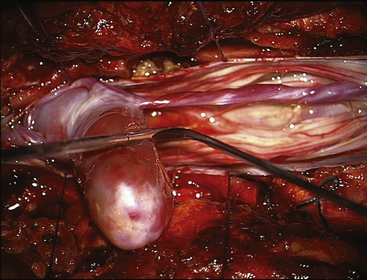
FIGURE 189-4 Operative view of the same patient as Figure 189-3 showing debulking of the superior pole of the tumor.

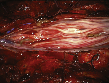
FIGURE 189-5 Operative view of the same patient as in Figures 189-3 and 189-4 demonstrating root preservation after tumor removal.
The upper part of the tumor sometimes grows into the lower conus, which has to be divided; the tumor does not usually invade the conus and can be separated from it. With large tumors, debulking is necessary; in such cases, mostly ependymomas, the tumor wall no longer exists and the tumor is always modified by intratumoral hemorrhage of different ages. The adjacent roots must be followed and preserved, bipolar coagulation must be used at a distance from them, and irrigation must be permanent. Of the sacral roots, the thinnest are the most exposed, and at least one is often injured during dissection.
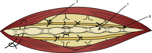
After complete tumor removal and careful hemostasis, the dura is closed. A dural graft is often necessary.7 The spine is reconstructed and the lumbar aponeurosis is attached to the spinous processes. It is recommended that one leave some aponeurosis attachment during the opening to facilitate this type of closure (Fig. 189-6). The subcutaneous fat and skin are closed as usual. The use of tissue glue is left to the preference of the surgeon. All the removed material is sent for histopathologic examination.

FIGURE 189-6 Fixation of the opened lamina with wires. 1, Spinous process; 6, supraspinous ligaments; 7, closure of aponeurosis with absorbable sutures.
After surgery the bladder catheter is left in situ for at least 3 days, and once the pain has grown less intense, the patient will be trained to perform self-catheterization until restoration of normal bladder function confirmed by urodynamic evaluation.
Pain is controlled with morphine in the early postoperative period. Specific treatment of deafferentation pain is instituted according to the type of discomfort described by the patient. Healing must be perfect by the time the patient is discharged to the rehabilitation facility.
An MRI evaluation must be scheduled for 3 months after surgery and serves as the reference in long-term follow-up of the patient.
Histologic Findings and their Consequences
Adult Pathology
Neurinomas (Schwannomas)
Schwannomas are the most common CETs, accounting for nearly 50% of our series. The mean age was 51 ±17 years. The time lapse between the first signs and the diagnosis is 2 years, and radiculopathy exists in nearly all cases, sometimes especially frequently during the night. Sphincter disturbance was present in 9% of patients, sexual dysfunction in 4.4%. Paravertebral spasm is found in 50% of patients and some degree of motor or sensory deficit in one third of patients.
Neurinomas can develop purely inside the spinal canal or simultaneously in the foramen (hourglass type), which can cause an enlargement of the neural foramen seen on neuroimaging. On CT scan and MRI there is homogeneous enhancement (Figs. 189-7 and 189-8). The tumor is well encapsulated, and complete removal can be achieved in most cases. Magnification allows identification of the rootlet bearing the tumor and preservation of the other roots (Fig. 189-9). The bearing rootlet is cut 1 cm above and 1 cm below the tumor, some very small satellite tumors or tumor prolongments being at times in close proximity to the tumor.

FIGURE 189-7 Sagittal T1-weighted magnetic resonance image (MRI) sequence without gadolinium of a benign neurinoma at the L2 level. The lesion is slightly hypointense to the conus medullaris.

Stay updated, free articles. Join our Telegram channel

Full access? Get Clinical Tree

