Figure 9-1 Left upper arm magnetic resonance imaging scan shows enlargement (arrow) of an approximately 6-cm segment of the median nerve in the distal arm, just anterior to the medial head of the triceps and approximately 2.2 cm from the skin level (A) and avid enhancement (arrow) of the focal segmental enlargement of the median nerve (B). The mass involves one fascicle of the left median nerve in the distal upper arm, which was purple in color and was very vascular (C).
The patient underwent removal of the mass, which involved only one fascicle of the left median nerve in the distal upper arm. Grossly, it was purple in color and was very vascular (see Fig. 9-1C). On paraffin sections, the nerve fascicles were found to be replaced by tumor, but there was no cellular atypia or inflammatory infiltration (Fig. 9-2A and B). There were no classifiable fibers on teased fibers. Myelinated fibers were absent on epoxy-embedded semithin sections. Immunohistochemical studies showed reactivity of blood vessels with CD-31, CD-34, and Factor VIII preparations (see Fig. 9-2C and D). This lesion was diagnosed as a glomangioma because of prominent proliferation of glomus cells and blood vessels but rare smooth muscle cells (Fig. 9-3).
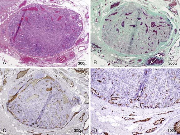
Figure 9-2 Paraffin sections show the nerve fascicles, which are replaced by tumor, but there is no cellular atypia or inflammatory infiltrations. Factor VIII shows reactivity with the vascular wall. H&E (A); Masson’s trichrome (B); Factor VIII (C) and (D).
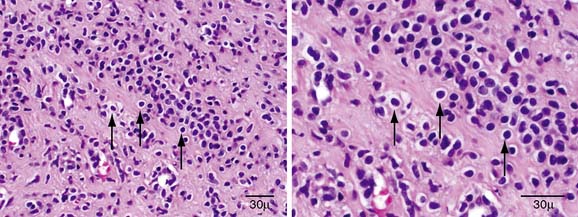
Figure 9-3 Paraffin sections show the prominent proliferation of glomus cells (arrows) and blood vessels but rare smooth muscle cells.
Related posts:
 Malignant Peripheral Nerve Sheath Tumor
Borrelia Neuropathy with Necrotizing Vasculitis
Adrenomyeloneuropathy Masquerading as Charcot-Marie-Tooth Disease
Focal Mononeuropathy Onset of Amyotrophic Lateral Sclerosis
A Case of Guillain-Barré Syndrome Associated with Anti-GD1b Immunoglobulin G Antibodies
Length-Related Axonal Loss in Neuropathy
Malignant Peripheral Nerve Sheath Tumor
Borrelia Neuropathy with Necrotizing Vasculitis
Adrenomyeloneuropathy Masquerading as Charcot-Marie-Tooth Disease
Focal Mononeuropathy Onset of Amyotrophic Lateral Sclerosis
A Case of Guillain-Barré Syndrome Associated with Anti-GD1b Immunoglobulin G Antibodies
Length-Related Axonal Loss in Neuropathy
Stay updated, free articles. Join our Telegram channel

Full access? Get Clinical Tree


