Fig. 53.1
Mucocele. Coronal T1-weighted precontrast MR image. The sphenoid is expanded and contains T1-hyperintense material. The normal pituitary gland is seen superior to the expanded sinus

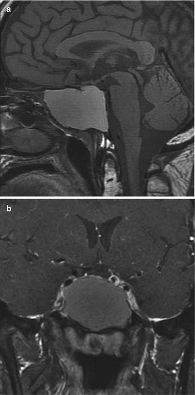
Fig. 53.2
Mucopyocele. (a) Sagittal T1-weighted pre-gadolinium MR image. (b) Coronal T1-weighted post-gadolinium image. The sphenoid is markedly expanded, containing T1 shortening material. The superior margin of the expanded sinus effaces the sella, and the pituitary gland is not clearly visible
53.3 Histopathology
Mucoceles are epithelial-lined cystic masses demonstrating an inflammatory reaction and mucinous material. Pseudostratified, ciliated, columnar epithelium is commonly noted, often with metaplasia in recurrent or chronic lesions.
53.4 Clinical and Surgical Management
The mainstay of treatment is endonasal transsphenoidal marsupialization and drainage.
Endoscopic endonasal management with the use of angled lenses is the gold standard surgical procedure, which facilitates maximal drainage of disease located in the lateral sphenoid recesses [16].
Antibiotics are typically used to augment therapy.
References
1.
2.
3.
Herman P, Lot G, Guichard JP, Marianowski R, Assayag M, Tran BA, Huy P. Mucocele of the sphenoid sinus: a late complication of transsphenoidal pituitary surgery. Ann Otol Rhinol Laryngol. 1998;107:765–8.CrossRefPubMed
Stay updated, free articles. Join our Telegram channel

Full access? Get Clinical Tree


