Chapter 111 Multilobar Resection and Hemispherectomy in Epilepsy Surgery∗
Multilobar Resection
The rationale of curative surgical procedures aiming at control of focal drug-resistant epilepsy is total inactivation (by resection or disconnection) of the epileptogenic zone (EZ), that is, the cortical region of onset and early spread of an ictal discharge.1 In this regard, the main efforts must be performed to correctly localize the EZ. Considering the dynamic features of an ictal discharge, and therefore its spatial–temporal structure,2 it is not surprising that in a number of patients the EZ may not be localized within the anatomic limits that mark the boundaries between the cerebral lobes.3 Moreover, the extension of an anatomic lesion that may correlate with seizures does not necessarily correspond to that of the EZ. Indeed, lesions limited to a single lobe may be associated with a multilobar EZ; in addition, anatomic abnormalities as wide as an entire hemisphere may be observed in patients with an EZ involving only a portion of a single lobe.
In patients with symptomatic focal epilepsy, the main problem consists of understanding the complex topographic and functional relationships between an anatomic (presumed to be epileptogenic) lesion and the EZ, and there is no general agreement concerning the strategy that best addresses this issue. Furthermore, in patients suffering from focal epilepsies with negative magnetic resonance imaging (MRI) (so-called cryptogenic epilepsies), the identification and spatial definition of the EZ must be based only on electrical and clinical findings.4
Preoperative Evaluation
Noninvasive Investigations
Collection of historical and clinical information is mandatory in the evaluation of candidates for surgical treatment of drug-resistant focal epilepsy. Age at seizure onset must be assessed, as well as seizure frequency and semeiology. Chronology of ictal symptoms must be accurately defined by carefully inquiring of both the patients and the witnesses about subjective manifestations and objective signs occurring during seizures. The possible occurrence of loss of contact must be assessed, as well as the presence of postictal deficits. This initial step may provide crucial clues to lateralization and/or gross localization of ictal onset.5
An interictal electroencephalogram (EEG) is helpful in determining the side and site of epileptiform and of slow-wave abnormalities. Long-term monitoring with scalp video-EEG recording, coupled with direct intensive surveillance of the patient allowing detailed ictal clinical examination, is mandatory when electroclinical correlates are needed.6
High-resolution MRI provides valuable anatomic information in these patients. The neuroradiologist must be aware of the electroclinical features of the patient to be evaluated to tailor the study to the individual case. In patients suspected to have a temporal lobe involvement, transverse and coronal slices are oriented parallel and perpendicular to the major hippocampal axis, respectively. Images are oriented according to the bicommissural line when electroclinical data suggest extratemporal epilepsy. Transverse double-echo, T2-weighted coronal turbo spin-echo (TSE), T2-weighted coronal TSE fluid-attenuated inversion recovery (FLAIR), and T1-weighted coronal inversion recovery are acquired in all patients, with additional sequences and slices obtained when needed.7 By these means, in more than 90% of the patients scheduled for surgery in our center (regardless of the site of resection), an anatomic lesion may be demonstrated.
In multilobar epilepsies, we often deal with highly functional areas and with cases presenting with altered anatomic patterns in eloquent regions (as are often encountered in malformations of cortical development). In these cases, functional MRI8 is particularly helpful in defining the limits of activated areas by, for instance, motor9 or speech tasks,10 thus allowing the surgeon to minimize the risks of unacceptable new neurologic deficits.11
The correlation among anatomic, EEG, and clinical data, supported by a full neuropsychologic evaluation, is then employed to formulate a coherent hypothesis as to the localization of the EZ. A high degree of correlation allows us to offer surgery to a large number of patients (currently about 70% in our center) without the need for further invasive investigations.
Invasive Investigations
When noninvasive investigations alone fail to correctly define the EZ, and/or when the EZ (or the anatomic lesion) is suspected to involve highly functional regions, invasive recording with intracranial electrodes is indicated. In many centers, subdural electrodes (arranged in strips or grids and placed through bur holes or craniotomy) are employed. In our center, stereotactic placement of intracerebral electrodes (stereoelectroencephalography, or SEEG) is preferred as the invasive EEG monitoring technique of choice, and it is required in approximately 30% of cases operated on. For each patient, the strategy of intracerebral electrode placement is tailored according to previously acquired clinical, electrical, and anatomic data. Examples of customized SEEG explorations are shown in Figs. 111-1 and 111-2. The technical details of this methodology have been reported elsewhere.12
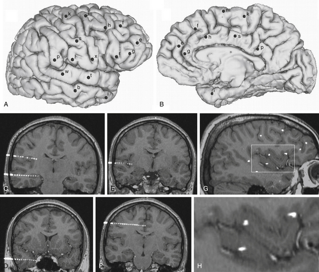
FIGURE 111-1 Example of an SEEG exploration. Coverage with intracerebral electrodes includes the right frontal and temporal lobes, as well as the rolandic and insular cortex. A brain cortical surface reconstruction of the lateral (A) and mesial (B) aspects of the right hemisphere was obtained from the 3D T1-weighted MRI structural images. (FreeSurfer software, http://surfer.nmr.mgh.harvard.edu.) Black spots, labeled by lowercase letters, indicate the surface impact points of each intracerebral electrode. C to H, Blended images of 3D T1-weighted MRI and coregistered postimplantation volumetric computed tomography, where single contacts of intracerebral electrodes are easily recognizable: electrodes b and l (C), electrode i (D), electrode t (E), electrode s (F), and a sagittal slice showing several contacts of different electrodes (G). H, The rectangular insert is magnified, where intracortical insular contacts of electrodes t, x, and r are shown.
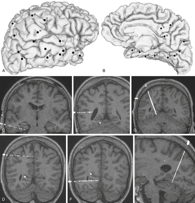
FIGURE 111-2 Example of an SEEG exploration. Coverage with intracerebral electrodes includes the right temporal, occipital, and parietal lobes. A brain cortical surface reconstruction of the lateral (A) and mesial (B) aspects of the right hemisphere was obtained from the 3D T1-weighted MRI structural images. (FreeSurfer software, http://surfer.nmr.mgh.harvard.edu.) Black spots, labeled by lowercase letters, indicate the surface impact points of each intracerebral electrode. C to H, Blended images of 3D T1-weighted MRI and coregistered postimplantation volumetric computed tomography, where single contacts of intracerebral electrodes are easily recognizable: electrode d and intrahippocampal contacts of electrode c (C), electrode p (D), electrode y (E), electrode o (F), and MRI slices coplanar with electrode k (G and H).
Surgical Decision-Making
The larger the epileptogenic cortex that needs to be removed, the harder the compromise that must be reached between the desire to cure each patient and the fear of creating new neurologic or neuropsychologic damage. The well-known concept of critical mass13 does apply to epilepsy surgery, and the decision on the extent of the EZ in multilobar partial epilepsies raises several particular diagnostic and therapeutic problems. In surgical planning, several questions can be considered that have no standard solutions and could have different answers depending on the characteristic of each patient and the attitude, feeling, and experience of the epilepsy surgery team. These issues concern the criteria for identifying the actual epileptogenic cortex. For instance, the following are challenging and not yet fully addressed questions: Should only the cortical areas that are affected by the ictal discharges at the onset of the seizures be removed? How long is the ictal onset? If the initial clinical symptom occurs several seconds after the onset of the electrical discharge, is removal of the symptomatogenic cortex mandatory,14 or will removal of only the initially affected cortical region be sufficient? What is the risk of removing the entire epileptogenic cortex? What kind of neurologic or neuropsychologic deficits can be induced? Are all of them predictable?
Anesthetic Considerations
Resective surgery for treatment of focal epilepsy does not require anesthetic approaches substantially differing from those commonly employed in other intracranial procedures. In our center, resections are performed under general anesthesia, with the patient positioned according to the site of surgery. Prophylactic antibiotics (1-2 g of cephamezine given intravenously) are given at anesthesia induction. Candidates for surgical treatment of severe partial epilepsy are commonly in optimal physical condition, and their interictal intracranial pressure (ICP) is not elevated.15 Careful anesthesiologic management (mostly hyperventilation, diuretics, or barbiturates) can be helpful when the volume of the exposed brain suddenly increases. This transitory increase in ICP may suggest the occurrence of an epileptic seizure,16 the clinical symptoms of which are masked by anesthesia. Except for this infrequent, transitory, and easily controlled incident, no other major intraoperative complications are usually expected in epileptic patients.
Surgical Technique
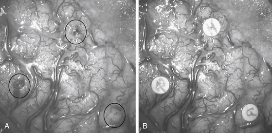
As compared to classic approaches for unilobar epilepsies, those adopted for multilobar excisions are tailored according to the site and extension of the region involved in the procedure. The supine position is usually preferred even when surgery is performed on the posterior portions of the hemisphere, with the operating table being tilted and the head rotated to obtain comfortable access to the surgical field. The prone position is used mainly when surgery involves the mesial aspects of the parietal or occipital lobes. Both skin incision and bone flap are designed to fully expose the region to be removed, as well as the surrounding cortical areas. In some instances, when a two-step procedure is likely, the surgeon must take into account the possible subsequent step when fashioning skin and bone flaps. In patients who have been previously evaluated by SEEG, the tracks of electrodes can be easily identified on the bone surface, and they are used as landmarks to plan the extent of the bone flap. While opening the dura mater, if an SEEG has been previously performed, care should be taken to separate the adhesions between the inner aspect of the dura and the cortical surface at the electrode entry points. Once the dura is opened, it is useful to match the vascular surgical anatomy with the stereoscopic angiograms obtained preoperatively, which provide an exquisite three-dimensional (3D) view of the sulcal and gyral pattern of the region.17 Careful inspection of the exposed cortex enables the surgeon to identify the entry points of intracerebral electrodes, which are used to draw the borders of resection (Fig. 111-3).
With the aid of the surgical microscope, resection is accomplished according to a few simple rules:
• Vessels that cross the region of resection but are tributaries of, or draining from, regions outside its borders must be identified on the stereoscopic angiograms, isolated, and left intact.
• Subpial dissection should be preferred, especially in mesial regions and along the main fissures, to protect vascular and extra-axial nervous structures (e.g., those in the prepeduncular cistern during mesial temporal removal).
• Suction and bipolar coagulation should be avoided to obtain as many unaltered tissue specimens as possible available for histopathologic evaluation.
Like hemispherectomy, which has progressively evolved into hemispherotomy (described later) to minimize the risks of hydrocephalus, hemosiderosis, and intraoperative blood losses, extended multilobar resections may be replaced with disconnective techniques. In our experience, disconnections have been particularly helpful in procedures involving the posterior quadrant of the hemisphere, when the EZ extends to the temporal, occipital, and parietal lobes. In these cases, we can employ a pure disconnective technique (Fig. 111-4) or otherwise combine resection with disconnection.18 Unlike other authors, when combining the two techniques, we prefer to remove the occipital and parietal lobes and to disconnect the temporal lobe, which may exert a sort of “push-up” effect if left in place, thus limiting residual brain dislocation into the resulting surgical cavity. Like other disconnective techniques, posterior hemispheric disconnections are not indicated if evolutive pathologies are suspected. A drawback of disconnective approaches is the limited availability of resected tissue, which may prevent a reliable etiologic diagnosis and restrict the access of basic scientists to pathologic specimens.
The anatomofunctional basis of morbidity in multilobar resections does not differ substantially from that of unilobar resections, and it strictly depends on the site of surgery. Morbidity may include transient motor impairment, akinesia and mutism after removal of the supplementary motor area,19 transient speech disturbances following resections involving the neocortex of the dominant temporal lobe, and contralateral superior quadrantoanopia in anterior temporal lobectomies. The risk of larger contralateral visual field defects is higher following resections in the posterior portion of the hemisphere. When removal is contiguous to the rolandic region, the risk of motor–sensory impairment is usually increased, and functional mapping by either intracerebral electrical stimulations at SEEG or functional MRI or intraoperative cortical and subcortical electrophysiologic evaluation is helpful to minimize the chances of unwanted postoperative deficits.
Results on seizures of multilobar surgery are far less gratifying if compared to other unilobar resections, either in our or in other centers18,20–26 (Tables 111-1 to 111-3). Slightly better results are observed in multilobar resections, including the temporal lobe. However, it must be stressed that, despite a complex epileptologic situation that could hardly be improved by further drug regimens, a substantial number of patients can be cured or improved (Engel’s27 classes I-III, approximately 80% in our series) by surgical treatment.
Table 111-1 Results on Seizures in Unilobar Resections∗
| Class | Patients |
|---|---|
| I | 438 (76.2%) |
| Ia+c | 365 (63.5%) |
| II | 52 (9.0%) |
| III | 45 (7.8%) |
| IV | 40 (7.0%) |
| Total patients | 575 (100%) |
Ia+c, completely seizure-free patients.
∗ Results are according to Engel’s (1987) classification.27
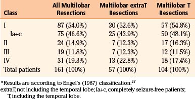
Table 111-3 Literature Reporting Seizure Outcome in Multilobar Resections (Publication Period 1998-2008)
| Described Population | Outcome: SF/Total (Only Multilobar) | |
|---|---|---|
| Elsharkawi et al.26 | 154 extratemporal resections | 7/15 (47%) |
| Daniel et al.18 | 13 patients with OPT epilepsy | 12/13 (92%) |
| Francione et al.22 | 10 pediatric patients with FCD | 1/2 (50%) |
| Kral et al.20 | 53 patients with FCD | 0/1 (0%) |
| Bernasconi et al.23 | 8 double cortex patients | 1/2 (50%) |
| Edwards et al.24 | 35 patients with MCD | 6/11 (55%) |
| Paolicchi et al.21 | 75 pediatric patients | 2/9 (22%) |
| Duchowny et al.25 | 31 pediatric patients | 2/5 (40%) |
FCD, focal cortical dysplasia; MCD, malformation of cortical development; SF, seizure-free; OPT, occipitoparietotemporal.
Illustrative Case
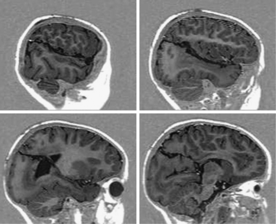
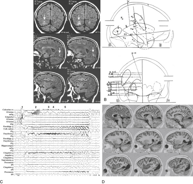
Scalp video-EEG disclosed slower background activity on the right hemisphere and interictal slow waves and spike and waves on the right temporo-occipital regions. Ictal EEG showed an early low-voltage fast activity on the right posterior leads, with late spread to the homologous contralateral regions. Brain MRI documented two separate lesions of possible ischemic nature in the right hemisphere, involving the basal occipital and the rolandic regions (Fig. 111-5A). SEEG investigation was performed to evaluate the relationships between the occipital anatomic lesion and the EZ, which could be suspected to involve also the parietal and temporal lobes on the basis of the electroclinical findings (Fig. 111-5B). No electroclinical data indicated the need to sample the rolandic lesion by intracerebral electrodes. Electroclinical correlations obtained during SEEG recordings localized the EZ in the inferior half of the occipital lobe (electrodes l and v) and in the posterior mesiobasal aspect of the temporal lobe (electrodes f and e) (Fig. 111-5C). Surgical resection of the EZ was performed (Fig. 111-5D), and histologic examination documented gliotic changes in the excised tissue corresponding to the MRI lesion. The patient was left with an expected and anticipated complete left-sided hemianopia, and she is seizure free since surgery after 3 years of follow-up.
< div class='tao-gold-member'>
Related posts:
 Chemotherapy for Brain Tumors
Current Surgical Management of High-Grade Gliomas
Endoscopic Endonasal Approach for Craniopharyngiomas
Revascularization Techniques in Pediatric Cerebrovascular Disorders
Surgical Management of Parasagittal and Convexity Meningiomas
Surgical Management of Major Skull Defects and Potential Complications
Chemotherapy for Brain Tumors
Current Surgical Management of High-Grade Gliomas
Endoscopic Endonasal Approach for Craniopharyngiomas
Revascularization Techniques in Pediatric Cerebrovascular Disorders
Surgical Management of Parasagittal and Convexity Meningiomas
Surgical Management of Major Skull Defects and Potential Complications
Stay updated, free articles. Join our Telegram channel

Full access? Get Clinical Tree


