Fig. 34.1
Optic glioma. (a) Axial T2-weighted image. (b) Coronal T2 inversion-recovery (IR) image. (c) Axial T1-weighted gadolinium-enhanced image. The optic nerves are diffusely enlarged bilaterally, with enhancement extending to the optic chiasm. There is dilatation of the optic sheaths, and the right globe is displaced anterolaterally

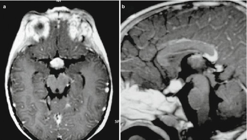
Fig. 34.2
Optic chiasm glioma in a 2-year-old patient with neurofibromatosis type 1. Axial (a) and sagittal (b) T1-weighted MR images after gadolinium administration show a homogeneously enhancing chiasmal tumor (Adapted with permission from Piccirilli et al. [15])
34.3 Histopathology
OPGs are comprised of various histopathological glioma subsets, including pilocytic astrocytoma, diffuse infiltrating astrocytoma, and pilomyxoid astrocytoma.
A majority of OPGs are pilocytic (WHO Grade I) astrocytomas, characterized by hypocellularity, glial fibrillary acidic protein (GFAP) immunoreactivity, and Rosenthal fibers (Figs. 34.3 and 34.4) [7]. Although mitoses are rare, microcystic degeneration is common.
A minority of OPGs are diffuse (WHO II) astrocytomas or pilomyxoid astrocytomas. The latter are characterized by myxoid vascular and perivascular cellular arrangements without Rosenthal fibers [8].
Although they are typically low-grade, indolent tumors, malignant transformation has been reported in 4–5 % of OPGs [9].

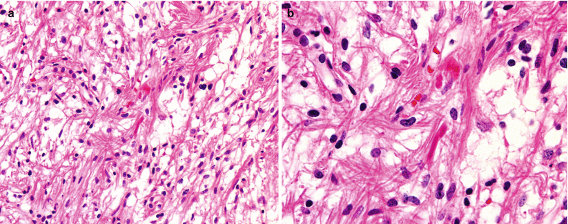
Fig. 34.3
Hypothalamic pilocytic astrocytoma. (a) Typical biphasic arrangement of compact, fibrillary pattern admixed with hypocellular and microcystic areas. (b) Numerous Rosenthal fibers are intermixed with the piloid cells

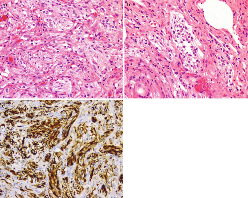
Fig. 34.4
Optic nerve pilocytic astrocytoma. Representative sections from a cystic tumor arising from the optic chiasm and involving the suprasellar region in a 13-month-old boy who presents with vomiting. The tumor shows spindle cells with coarse fibrillar processes and minimal nuclear pleomorphism arranged in fascicles (a). Rosenthal fibers are not prominent. A focal biphasic pattern is present (b). Immunohistochemical staining for glial fibrillary acidic protein (GFAP) is strongly positive (c)
34.4 Clinical and Surgical Management
Multimodality algorithms for OPGs include surgical resection versus biopsy, chemotherapy, and radiation treatments. Many clinicians advocate the use of early chemotherapy, reserving radiation treatment for as late as possible in children [10, 11].
Surgical treatment for OPGs is rarely curative and typically is limited to biopsies or minimal debulking. Many authors support empiric treatment with chemotherapy based on clinical and imaging diagnosis alone, with surgery reserved for refractory cases [12]. Early surgery may be indicated for large tumors causing hydrocephalus or for those with unilateral optic nerve involvement [7].
Stay updated, free articles. Join our Telegram channel

Full access? Get Clinical Tree


