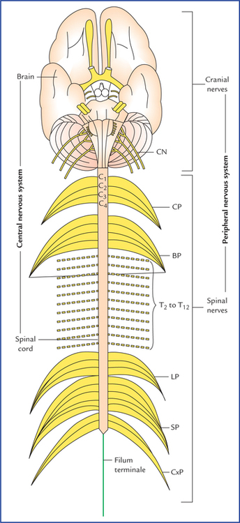
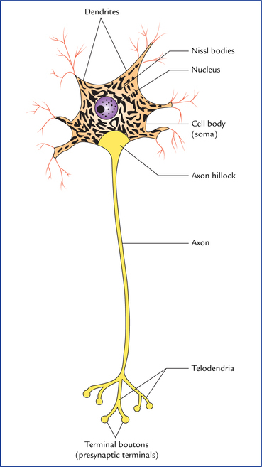
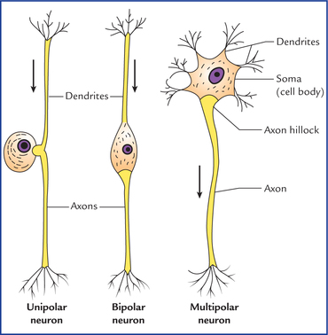
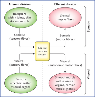
2 Neuroanatomy is the study of the nervous system. The nervous system is the most complex, widely investigated and least understood system in the body. It along with endocrine system regulates the functions of all other systems of the body. Hence nervous system is also called master system of the body. The functions of the nervous system include: • Reception of sensory stimuli from internal and external environments. • Integration of sensory information. • Coordination and control of voluntary and involuntary activities of the body. • Assimilation of experiences, a requisite to memory, learning and intelligence. • Storage of experiences to establish pattern of responses in future, based on prior experience. The mechanism of functioning of the nervous system is as follows: The sensory stimuli (afferent impulses) received from inside or outside the body are correlated within the nervous system and then coordinated motor response (motor impulses) is sent to the effector organs (muscles, glands, etc.) so that they work harmoniously for the well-being of the individual (Flowchart 2.1). Anatomically the nervous system is divided into two parts, the central nervous system and the peripheral nervous system (Fig. 2.1). fig 2.1 Anatomical divisions of the nervous system. The central nervous system consists of brain and spinal cord. The peripheral nervous system consists of cranial nerves which arise from the brain, and spinal nerves which arise from the spinal cord. (CP = cervical plexus, BP = brachial plexus, LP = lumbar plexus, SP = sacral plexus, CxP = coccygeal plexus, CN = cranial nerves.) • The central nervous system (CNS) consists of brain and spinal cord. The brain is located within the cranial cavity and the spinal cord within the vertebral canal. The CNS is responsible for integrating, processing, and coordinating sensory data, and giving appropriate motor commands. It is also the seat of higher functions such as intelligence, memory, learning, and emotions. • The peripheral nervous system (PNS) includes all the neural tissues outside the CNS, such as 12 pairs of cranial nerves, 31 pairs of spinal nerves, and ganglia associated with cranial and spinal nerves. The PNS provides sensory information to the CNS and carries its motor commands to the peripheral tissues and systems. Functionally also the nervous system is divided into two parts, the afferent division and the efferent division (Fig. 2.2). • The afferent division brings sensory information to the CNS. • The efferent division carries motor commands to the muscles and glands. – The somatic nervous system (SNS) provides the voluntary control over the skeletal muscle contraction. – The autonomic nervous system (ANS) innervates involuntary structures, such as heart, smooth muscle and glands and thus provides an involuntary regulation of smooth muscle, cardiac muscle, and glandular activity. The highly specialized and complex nervous system consists of only two principal categories of cells, (a) neurons, and (b) neuroglia. • Neurons form the basic structural and functional units of the nervous system. They are excitable cells which are specialized for reception of stimuli and the conduction of nerve impulses. • Neuroglia or glial cells are supportive cells that support the neurons both structurally and functionally. The neu-roglia are five times more abundant than the neurons and account for more than half of the weight of the brain. The typical neuron has a single long process called axon and many short processes called dendrites (Fig. 2.3). Fig. 2.3 A neuron. Note that Nissl substance is distributed throughout the cytoplasm of the cell body except in the region close to axon called axon hillock. It extends into the dendrites but is locking in the axon. The neurons exhibit considerable diversity in form and function. Therefore, they are classified structurally as well as functionally. • Pseudounipolar neurons. These neurons possess oval or rounded cell body. A single process emerges from the cell body and after a short convoluted course bifurcates at a T-junction into peripheral and central processes. They are called pseudounipolar neurons because it is thought that the two processes of the bipolar neurons, during the process of differentiation, are approximated and finally fused near the cell body to form a single process. Thus, it appears that the neurons possess a single process bifurcating in a T-shaped manner, a short distance from the cell body. Such neurons are found in dorsal root ganglia of spinal nerves and sensory ganglia of some cranial nerves. Fig. 2.4 Three basic morphological types of neurons. The arrows indicate the usual direction of impulse transmission. • Bipolar neurons. They possess spindle-shaped cell body, from each end of which a single neurite (process) emerges. Thus, bipolar neurons have two processes, one dendrite and one axon, with the soma between them. Such neurons are found in olfactory epithelium of nasal cavity, retina of eyeball and sensory ganglia of cochlear and vestibular nerves. • Multipolar neurons. Have multipolar cell body from which emerges several dendrites and a single axon. Most of the neurons in the body especially those in CNS belong to this category. For example, all the motor neurons that control skeletal muscles are multipolar neurons. • Golgi type I neurons. These neurons have long axons that may be one metre long in extreme cases and connect different parts of the nervous system. The axons of these neurons form the long fibre tracts of the brain and spinal cord, and the nerve fibres of the peripheral nerves. The pyramidal cells of the cerebral cortex, Purkinje cells of the cerebellum and motor anterior horn cells of spinal cord are Golgi type I neurons. Their dendrites are short and numerous. • Golgi type II neurons (microneurons). Axons of these neurons are morphologically similar to that of dendrites. This gives these cells a star-shaped appearance. They establish synaptic contacts with large number of neurons in their neighbourhood. They are found in large numbers in cerebral cortex, cere-bellar cortex and in the retina. Table 2.1 summarizes the morphological (anatomical) classification of neurons. Table 2.1 Morphological classification of neurons They carry impulses from the receptor organs to the CNS. In relation to the general sensory pathways, they are classified into three types: 1. Primary sensory neurons: The cell bodies of these neurons lie outside the CNS except those of mesencephalic nucleus of fifth cranial nerve which lie within the CNS. 2. Secondary sensory neurons: The cell bodies of these neurons lie in the CNS. 3. Tertiary sensory neurons: The cell bodies of these neurons lie in the thalamus. For details see Chapter 17. In the somatic nervous system they are divided into two types: 1. Upper motor neurons have their cell bodies located in the cerebral hemisphere, viz. motor area of the cerebral cortex. They form the descending pathways of the brain and synapse with the motor neurons of the cranial nerve nuclei in the brainstem and motor neurons of the spinal nerves in the anterior horns of the spinal cord. The upper motor neurons are involved in the voluntary control of muscular activity. 2. Lower motor neurons have their cell bodies located in the brainstem and spinal cord.
Organization and Functions of the Nervous System
Divisions of Nervous System
Anatomical

Functional
Cellular Organization of the Nervous System
Neurons (Neuro, Nerve)

Classification of neurons (types of neurons)
Anatomical (morphological) classification
According to polarity (Fig. 2.4)

According to relative lengths of axons and dendrites
Morphology
Location and example
According to polarity
• Unipolar/pseudounipolar
Posterior root ganglia of spinal nerves, sensory ganglia of cranial nerves
• Bipolar
Olfactory epithelium, retina, sensory ganglia of cochlear and vestibular nerves
• Multipolar
Central nervous system (motor cells forming fibre tracts of brain and spinal cord and peripheral nerves), autonomic ganglia
According to size of nerve fibre
• Golgi type I
Pyramidal cells of cerebral cortex, Purkinje cells of cerebellum, anterior horn cells of spinal cord
• Golgi type II
Cerebral cortex, cerebellar cortex (stellate cells forming synaptic contacts with other neighbouring neurons)
Functional classification
Types of sensory neurons
Types of motor neurons
![]()
Stay updated, free articles. Join our Telegram channel

Full access? Get Clinical Tree


