Fig. 18.1
The 9-year-old female patient with SGS. The patient had a previous growing rod instrumentation to treat pelvic obliquity (a, b), however, developed lordosis and significant decompensation with a right trunk shift. (c, d) show images following initial growing rod surgery. In postoperative follow-up, this patient has had three lengthenings in 3 years with a total increase of 4 cm in T1–S1 length (e, f). Complications since initial surgery have included distal screw breakage and rod erosion that required successful revisions
18.2 Ehlers–Danlos Syndrome
Ehlers–Danlos syndrome (EDS) is a class of connective tissue disorder caused by defects in collagen synthesis. It is characterized by distensible and thin skin, easy bruising, hyperextensible joints, facial features, and severe arterial complications. The eye, gastrointestinal, respiratory, and cardiovascular systems can also be affected.
18.2.1 Etiology/Genetics
EDS is not a homogeneous disorder and can be thought of as a group of related entities that share, to varying degrees, the same complex of physical anomalies. Therefore, various subclassifications exist with different clinical presentations and different genetic mutations. According to the Villefranche classification, Type VI is characterized by progressive infantile scoliosis. It is inherited in an autosomal recessive fashion with mutation in PLOD gene (encoding lysyl hydroxylase important in collagen cross-linking) [9].
18.2.2 Skeletal/Spine Manifestations
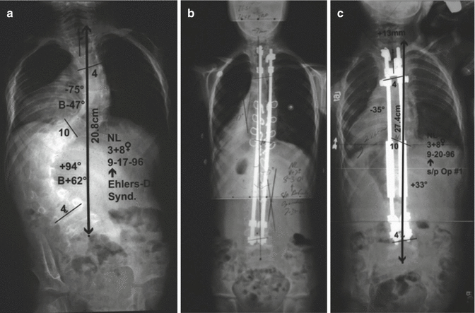
Skeletal manifestations include developmental dysplasia of the hip, club foot, pes planus, joint hypermobility and dislocation, generalized ligamentous laxity, and scoliosis [9]. Kyphoscoliosis is a hallmark feature of type VI EDS; however, scoliosis also often presents at an early age in patients with other classes of EDS, most notably Types I, II, and III [10] (Fig. 18.2). Osseous fragility is often seen.


Fig. 18.2
The 3-year-old female with EDS with preoperative curve over 90° (a) underwent placement of growing rods (b). The patient later went on to successful posterior fusion at the age of 8 (c) with curve at last follow-up of 25° (Case courtesy of Marc A. Asher, MD)
18.2.3 Surgical Treatment and Complications
With scoliosis surgery, it is important to keep in mind the vascular fragility that is inherent in this disease. Although spine surgery in MFS can be associated with increased bleeding, anterior approaches to the spine should be avoided when possible in EDS because such surgery can be catastrophic involving large arteries and veins. Akpinar’s review of five cases with Type VI EDS who underwent surgical treatment of scoliosis had two cases of vascular complications during the anterior approach, one involved avulsion of the segmental arteries from the lower aorta and common iliac vein requiring gortex graft repair [10]. Vogel et al. also reported major vascular complication in one out of four patients associated with the anterior approach due to the inherent vascular fragility in EDS patients. They also reported major neurological complications including permanent paraplegia in two patients [11].
Growing procedures in EDS patients, if begun early enough, may allow posterior-only approaches so that the patient never requires an anterior procedure or a complex posterior osteotomy. Measures such as hypotensive anesthesia and careful dissection of segmental arteries are advised [10]. Recently, a report of using Factor VIIa to help control massive bleeding following spontaneous large vessel rupture in Type IV EDS has been published [12].
18.3 Prader–Willi Syndrome
Prader–Willi syndrome (PWS) is characterized by early hypotonia, developmental and motor delay, small hands and feet, and later hyperphagia resulting in massive obesity.
18.3.1 Etiology/Genetics
PWS is caused by a lack of paternal expression of a region of chromosome 15. Pituitary dysfunction results in many generalized (listed earlier) and orthopedic manifestations [13].
18.3.2 Natural History/Skeletal Manifestations
Orthopedic manifestations include growth retardation, hip dysplasia, and scoliosis. Scoliosis is seen in 66 % of patients with PWS by time of skeletal maturity, according to longitudinal study by Odent et al. [14]. Its onset is often in the infantile or juvenile period with a mean age of onset 10.2 ± 6.2 years [14]. Increased body mass index (BMI) was a risk factor for developing associated kyphotic deformity which led to a higher likelihood of surgical treatment. Annual systematic clinical examination for scoliosis is recommended.
18.3.3 Spinal Deformity Treatment and Complications
Orthoses have a role if body habitus does not prohibit it. Administration of human growth hormone (HGH) has been shown to aid in the management of many aspects of this condition [13]. Initially, there was concern that HGH may increase the prevalence and severity of scoliosis [13]. However, it appears to help control many aspects of the disease and does not increase the incidence or severity of scoliosis [14]. In fact, a study out of South Korea purports that in their clinical series preoperative treatment with HGH before surgical fixation may reduce postoperative complications [15]. Massive obesity is now rarely seen in PWS since the advent of HGH treatment.
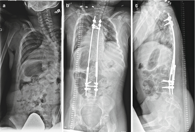
Treatment of scoliosis in this condition should follow usual clinical guidelines. If curve size increases beyond orthotic range in the early juvenile period, there may be a role for growth-guiding surgery (Fig. 18.3). At the time of any surgical procedure, monitoring for sleep apnea is important. Other relevant important perioperative considerations are that these patients have higher likelihood of osteopenia, depression, and diminished pain sensitivity [16]. Proximal junctional kyphosis with acute cord stenosis has been reported after spine surgery in PWS.


Fig. 18.3
The 9-year-old male with PWS. The patient had a collapsing 90° scoliosis that progressed despite attempted bracing (a). Growing rods were inserted and b and c display images after second growing rod surgery with a Cobb angle reduced to 36°
18.4 Rett Syndrome
Rett syndrome (RS) is a rare progressive neuromuscular disorders first described a generation ago by Austrian physician Andreas Rett. It is often confused with cerebral palsy.
18.4.1 Etiology/Genetics
18.4.2 Presentation
Virtually all patients are female, and manifest stereotypic hand movements, little to no expressive language, seizures, and a neurological picture combining dystonia and spasticity.
Stay updated, free articles. Join our Telegram channel

Full access? Get Clinical Tree


