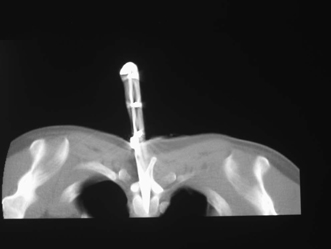
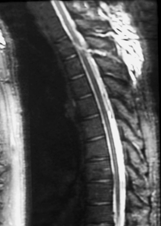
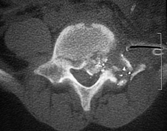
21 Penetrating Spine Trauma Michael D. Martin and Christopher E. Wolfla Penetrating spine trauma encompasses injury caused by firearms, both military and civilian, as well as foreign bodies, including knives and a myriad of other implements. It is in essence largely a social problem, and perhaps summarized best in a quote from the Lancet, ca. 1962: “In an instant-and often for a negligible reason-an otherwise healthy man is incapacitated, either permanently or for many months.”1 Reports vary widely as to the proper management of such injuries, but certain guiding principles can be gleaned from the available data. This chapter distills what is known about these injuries, as well as provides a logical approach to the management of penetrating spinal trauma. Finally, we will discuss the outcome of these often devastating injuries with respect to neurological recovery. The incidence of spinal cord injury in penetrating neck injuries is between 3.7% and 15.0%.2,3 The average age of victims of civilian-type gunshot wounds in one series was ~32 years of age, with 89% of the victims being male.4 Not surprisingly, the same series of 92 patients revealed that the thoracic region was the most often injured (59%), followed by the cervical spine (31%), and finally the lumbar spine (10%). Seventy-five percent of these had complete injuries, whereas 25% had incomplete injuries. With regard to military gunshot wounds, a large review of injuries from World War II revealed that most wounds were located near the midline and at the cervicothoracic junction.5 Literature from the Vietnam conflict cited the thoracic spine as the most common location.6 Spinal injury from stab wounds also occurred more frequently in males (84% compared with 16% females in a large series).7 A very interesting series from South Africa reported this injury was caused most commonly by knives (84.2%), though a surprising number of implements may be involved.7 Here again the thoracic region was the most commonly involved (63.8%). In the entire series, 20.9% of the patients suffered complete spinal cord injuries. In all types of penetrating spinal injury, initial history and physical examination are important in guiding both the need for further investigation and the proper type of treatment. History must be obtained to best delineate the probable mechanism by which the spinal cord has been injured. Nowhere is this more evident than in the difference between a wound from a civilian versus a military firearm. Wounding patterns differ between these two types of weapons because of ballistics.8,9 As a bullet passes through tissue a sonic pressure wave precedes the projectile without causing injury in and of itself.9 As expected, bullets are rapidly slowed as they enter tissue, and this rapid deceleration creates a temporary cavity in the tissue. This process is often referred to as cavitation.9 The amount of cavitation is related to the velocity of the projectile involved, with wounding capacity quadrupling as velocity is doubled.8 Physical examination is important in all patients, and documentation of motor and sensory levels is important in any spinal cord injury. By convention, spinal cord injuries are identified by the lowest level of antigravity motor function. Assessment of entrance and exit wounds can be useful in determining trajectory, which has been shown to be an important factor in the severity of injury suffered.10 A statistically significant difference has been found in the degree of spinal cord injury suffered by those in whom a bullet has traversed the spinal canal and those in whom it did not (88% complete injuries if the bullet traversed the canal, 78% incomplete if it did not).10 Stab wounds most often (63.8%) involve the thoracic spine, followed by the cervical spine (29.6%) and finally the lumbar region (6.7%). Complete cord injury also occurred in a higher percentage of patients stabbed in the thoracic spine (24%) than in the cervical (15.8%) or lumbar spine (10%).7 In this type of nonmissile penetrating trauma, the bony elements of the spine seem to deflect injuries to either side of the midline, decreasing the chance of complete cord injury1,11 (Fig. 21-1). The implement used may directly injure the spinal cord, may injure arterial supply or venous drainage, or may cause a contre-coup type of cord contusion.1,7 This may lead to injury patterns that do not follow the classic Brown-Séquard pattern, even in the case of a knife causing anatomical hemisection of the cord.7 Laminar fractures have been reported if the instrument used was of sufficient size and mass.1 Initial imaging should include complete spine x-rays, computed tomography (CT), and magnetic resonance imaging (MRI) when available and clinically feasible.12,13 Some have suggested that it is unnecessary to use cervical spine immobilization in fully conscious patients with isolated penetrating trauma,14 though it must be remembered that cervical spine fractures in gunshot wounds to the neck may occur in 14.6% to 43.0% of patients.15,16 Cervical instability is a possible, albeit rare, sequela of penetrating neck trauma.17 Although MRI is an important tool in spinal cord injury, quality may be decreased by ferromagnetic artifact from foreign body residue.11 MRI is not recommended in the case of a known retained metallic foreign body. Penetrating trauma may cause neurological deficit from epidural or subdural hematoma, disk herniation, foreign bodies within the spinal canal, or displaced bony elements of the spinal column.13 Cord contusions may appear as high-signal-intensity areas on proton density-weighted and T2-weighted MRI, whereas T2-weighted images are probably the best sequence for evaluating cord edema.13 Acute or subacute hemorrhage can be represented by a focus of low-signal intensity on T2-weighted and proton density-weighted images, or an area of high signal intensity on T1-weighted, T2-weighted, and proton density-weighted sequences.13 Intramedullary knife tracts are best demonstrated as high-signal-intensity lesions on T2-weighted and proton density-weighted images13 (Fig. 21-2). Subdural hematomas usually demonstrate a concave surface facing the cord and a convex surface toward the adjacent vertebral body, whereas epidural hematomas are often biconvex (as in intracranial epidural hematomas).13 Figure 21-1 Axial computed tomographic scan demonstrating a knife in the spinal canal. It has been deflected from the midline somewhat by the bony spinous process. A review of the literature finds that the incidence of vertebral artery injury in penetrating cervical trauma is 1.0% to 8.0%.2,18–20 In most cases, physical examination and CT of the bony elements of the cervical spine provide reliable evaluation of vascular insult in the neck and should be used to guide the decision for further vascular imaging16,21–24 (Fig. 21-3). Injury may include occlusion, arteriovenous fistula, intimal tear, or pseudoaneurysm.25,26 Up to 20% of patients may have no signs at all,20 and vertebral artery injury in the absence of cervical spine fracture is rare.16 Most authors advocate angiography as the standard evaluation tool for suspected vertebral artery injury.27,28 Magnetic resonance angiography (MRA) and computed tomographic angiography (CTA) hold the promise of noninvasive diagnosis, but their usefulness in evaluating vertebral artery trauma has not been established.29,30 CT and CTA are useful in detecting other indirect signs of vascular injury, including bullet and bone fragments less than 5 mm from a major vessel, injury path through a vessel, or hematoma around the vessel.29 Direct signs of vascular injury visible on CTA include changes in vessel caliber, irregularities in the vessel wall, extravasation of contrast, and lack of enhancement.29 MRA has a reported specificity of 98% to 100% but a sensitivity of 20% to 60% (depending on the sequence used) in detecting vertebral artery injury.29 MRA has lower resolution than arteriography and at present is not recommended over arteriography for diagnosing vertebral artery injury.31 Figure 21-2 T2-weighted magnetic resonance image of the spine following knife injury. The knife tract is hyperintense on this sequence. Figure 21-3 Axial computed tomographic scan following gunshot wound to the spine demonstrating disruption of bony elements. Vertebral arteriovenous fistula (AVF) is a rare complication of penetrating spine or neck trauma and may develop some time after the initial injury.32 The most common symptom is tinnitus, present in 39% of patients in one series.33 Other symptoms include headache, vertigo, diplopia, cervical neuralgia, or neck mass.32,33 Roughly 41% of patients have no neurological symptoms and present with only a cervical bruit. Heart failure is one possible sequela of any AVF, including those arising from the vertebral artery.33,34 Another rare presentation is cervical cord or nerve root compression from draining veins arising from the AVF.33,35 Headache has been noted as a sequela of gunshot wounds to the spine.36 A rare but interesting late-onset symptom is that of plumbism from retained bullet fragments in the disk space, which should resolve with removal of the fragments.37 Others have reported osteomyelitis or sepsis following gunshot wounds to the spine that traversed the gastrointestinal tract, but larger series suggest that this is a rare entity.38–41 Treatment of victims of penetrating spinal trauma is dependent on both the mechanism of injury and the patient’s early postinjury course. Although currently standard practice for victims of other types of spinal cord injury, methyl-prednisolone increases complications but does not improve outcomes in patients who are victims of penetrating spinal trauma.42,43 Some series have recommended aggressive surgical treatment for all gunshot victims,44 and one larger series showed improvement after bullet removal only in lesions at T12 or below.40 Other series have shown, however, that operating on all victims of civilian gunshot wounds conveys no significant improvement over conservative management and may increase the risk of infection, cerebrospinal fluid leakage, pseudomeningocele, and spinal instability.4,43,45–47 Indications for surgical intervention in civilian gunshot wounds to the spine therefore include progressive neurological deficit and persistent spinal fluid leakage, although most authors feel that these are rare entities.4,43,46,48 Although technically difficult, surgery may be beneficial in the case of incomplete injury with evidence of continued neural compression.43 A smaller series of patients with incomplete injuries of the cauda equina showed a worse outcome with surgery (47% improvement) compared with conservative management (71% improvement).49 Victims of shotgun injuries to the spinal cord have demonstrated no significant improvement following laminectomy50 and have an overall increased mortality when compared with other gunshot victims.51 Experience in the military literature has been quite different. Although some studies have found results similar to the civilian data,52,53 many authors advocate an aggressive approach to management of penetrating spinal trauma from military (i.e., high-velocity) weapons.54–57 Laminectomy, foreign body removal, and dural repair, if possible, in all patients with neurological deficit and without irrefutable evidence of complete anatomical transection has been shown in the military literature to provide some measure of recovery in 47.6% to 52.4% of patients.54,56,57 Stab wounds inflicted by knives or other foreign bodies are best treated with the same general approach as gunshot wounds. Indications for surgery in the case of nonmissile penetrating trauma include retained foreign body material, persistent cerebrospinal fluid leakage, and development of sepsis from a sinus tract or epidural abscess.1,7 In a very large series spinal fluid leakage was encountered in only 4% of cases, and almost always resolved spontaneously. The development of sepsis did not occur.7 Other authors advocate routine exploration for all nonmissile penetrating trauma, albeit from a small series of patients.58 Open surgical reconstruction of the injured vertebral artery has been advocated and described by some authors,59 with a mortality of 4.7% to 22.0%.60,61 The development of effective endovascular techniques, however, has led to their use for the treatment of most injuries to the vertebral artery, including AVF and pseudoaneurysm.33,62–65 Emergency intervention is sometimes indicated due to active bleeding or hemodynamic instability.64 Attention should be paid to the patency of the contralateral vertebral artery as well as the location of any arteries feeding AVFs66 because a patent artery on the opposite side is a good indicator of the safety of ligation of the injured vertebral artery.67 Pseudoa-neurysms should be treated with anticoagulation if they are not occluded, and endovascular or surgical treatment if they persist.25 A large series comparing surgically versus conservatively managed penetrating trauma patients found no differences between the two groups in terms of neurological outcome.47 Those with complete injuries from gunshot wounds showed mild improvement in 13% to 15% and worsening of their deficit in 3% to 6%. Incomplete injuries improved in 40% to 58% of patients and worsened in 18% to 20% of cases. Similar results were seen in stab wounds. Overall morbidity from penetrating spinal injuries in the military literature has decreased since the early twentieth century and was reported as 2.3% in one paper from the Vietnam era.52 A large body of data from the Korean conflict (in which almost all patients had operations) divided outcome data by the level of injury.56 All of those with incomplete lesions of the cervical spine had some recovery following laminectomy (28.6% full recovery, 71.4% partial recovery). Thirty-five percent of those with complete cervical injuries showed no improvement, 60% had partial recovery, and 5% made a complete recovery. Incomplete lesions in the thoracic region were similar, with 20% achieving full recovery and 80% partial recovery, but patients with complete lesions in the thoracic region recovered function only 9% of the time (90% partial recovery, 10% full recovery). Partial injuries to the lumbar spine resulted in full recovery only 14.2% of the time, and complete injuries in this region showed some recovery in 18.8% of patients. A review of 450 cases of stab wounds to the spine demonstrated that recovery was good (meaning able to ambulate with minimal support) in 65.6% of patients.7 The vast majority of the patients (95.6%) in this series were not treated surgically. The authors went on to say that 17.1% of their patients made a “fair” recovery (walking with moderate assistance) and 17.3% made no functional recovery. Penetrating spinal trauma can cause devastating injury to otherwise healthy and most often young individuals. Neurosurgeons must use the clinical history, when available, as well as detailed physical examination and appropriate imaging to guide treatment and evaluate for other injuries such as vascular deformation. Although intervention may help patients who suffer wounds from high-velocity weapons or those resulting in spinal instability or vascular insult, the majority of patients seen in the urban trauma center will not require operative intervention. Perhaps the advances in spinal cord rehabilitation and research will add to the somewhat limited armamentarium with which neurosurgeons currently treat these devastating injuries. 1. Lipschitz R, Block J. Stab wounds of the spinal cord. Lancet 1962;28:169–172 4. Kupcha PC, An HS, Cotler JM. Gunshot wounds to the cervical spine. Spine 1990;15:1058–1063 6. Jacobson SA, Bors E. Spinal cord injury in Vietnamese combat. Paraplegia 1970;7:263–281 11. Takhtani D, Melhem ER. MR imaging in cervical spine trauma. Clin Sports Med 2002;21:49–75 20. Roberts LH, Demetriades D. Vertebral artery injuries. Surg Clin North Am 2001;81:1345–1356 26. Mwipatayi BP, Jeffery P, Beningfield SJ, et al. Management of extracranial vertebral artery injuries. Eur J Vasc Endovasc Surg 2004;27:157–162 55. Haynes W. Acute war wounds of the spinal cord. Am J Surg 1946;72:424–433 60. Demetriades D, Stewart M. Penetrating injuries of the neck. Ann R Coll Surg Engl 1985;67:71–74 61. Reid JD, Weigelt JA. Forty-three cases of vertebral artery trauma. J Trauma 1988;28:1007–1012
Introduction and Epidemiology
Initial Evaluation and Imaging


Treatment
Neurological Outcomes
Conclusion
References
< div class='tao-gold-member'>
Penetrating Spine Trauma
Only gold members can continue reading. Log In or Register to continue

Full access? Get Clinical Tree


