Fig. 35.1
Pituicytoma. (a) Sagittal T1-weighted precontrast image. (b) Coronal T1-weighted gadolinium-enhanced image. There is a hypoenhancing lesion in the right aspect of the sella, without suprasellar extension. Remodeling of the right sellar floor is also seen
35.3 Histopathology
Pituicytoma is classified as a WHO Grade I spindle cell astrocytic tumor originating in the neurohypophysis or infundibulum.
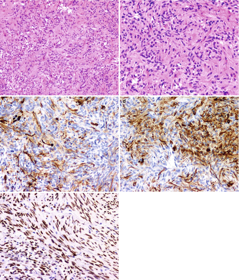
Pituicytoma consists of plump spindle cells with slightly pleomorphic nuclei, pinpoint nucleoli, and rare or absent mitoses [2].
Specimens are generally positive for S100 and vimentin immunostaining and are nonreactive for synaptophysin and neurofilament protein [2]. GFAP staining may be focally or diffusely positive (Fig. 35.2).
Most significantly, pituicytomas show strong nuclear TTF-1 immunoreacitivity (Fig. 35.2e).
The MIB-1 labeling index is reported to be low (1–2 %) in most cases [14].
Pituicytoma can be differentiated from normal neurohypophysis, which contains axons and Herring bodies [2].
Pituicytomas also can be differentiated from pilocytic astrocytomas, which have Rosenthal fibers and granular bodies [15].
Ultrastructural analysis shows absence of secretory granules in pituicytoma [16].
Sellar ependymoma is a rare lesion with classic ependymal cell findings; it is thought to arise from ependymal pituicytes [10].

Fig. 35.2
Pituicytoma. Pituicytomas are characterized by elongated fibrillary cells arranged in a fascicular pattern. The cells have a piloid, fibrillary appearance similar to pilocytic astrocytomas (a, b, H&E stain). Tumor cells are strongly immunoreactive for GFAP (c) and vimentin (d). Thyroid transcription factor 1 (TTF-1) nuclear immunoreactivity is strongly positive in pituicytomas (e) and seems to confirm an origin from the intrinsic glial cell of the pituitary, the pituicyte
35.4 Clinical and Surgical Management
Transsphenoidal surgery is typically an effective approach for intrasellar pituicytoma. For purely suprasellar pituicytoma, craniotomy may be preferred, as pituicytomas have been reported to be highly vascular, firm tumors that may result in considerable intraoperative bleeding [17].
Aggressive surgical management is recommended when feasible, although a gross total resection often cannot be achieved safely.
The vascular supply of pituicytomas has been shown to derive in part from the superior hypophyseal arteries, and selective embolization prior to surgery has been suggested if the diagnosis is known ahead of time [3, 17].
Although no large series with long-term follow-up are available, gross total resection has been associated with long-term recurrence-free follow-up. Recurrence appears to be common following subtotal surgical resection, occurring in roughly 39 % of patients [9, 18].
Adjunctive radiation therapy may have been beneficial in anecdotal cases, although no clear clinical benefit has been shown [9].
Hormone replacement therapy for hypopituitarism is often required after treatment.
References
1.
Louis DN, Ohgaki H, Wiestler OD, Cavenee WK, Burger PC, Jouvet A, et al. The 2007 WHO classification of tumours of the central nervous system. Acta Neuropathol. 2007;114:97–109.PubMedCentralCrossRefPubMed
Stay updated, free articles. Join our Telegram channel

Full access? Get Clinical Tree


