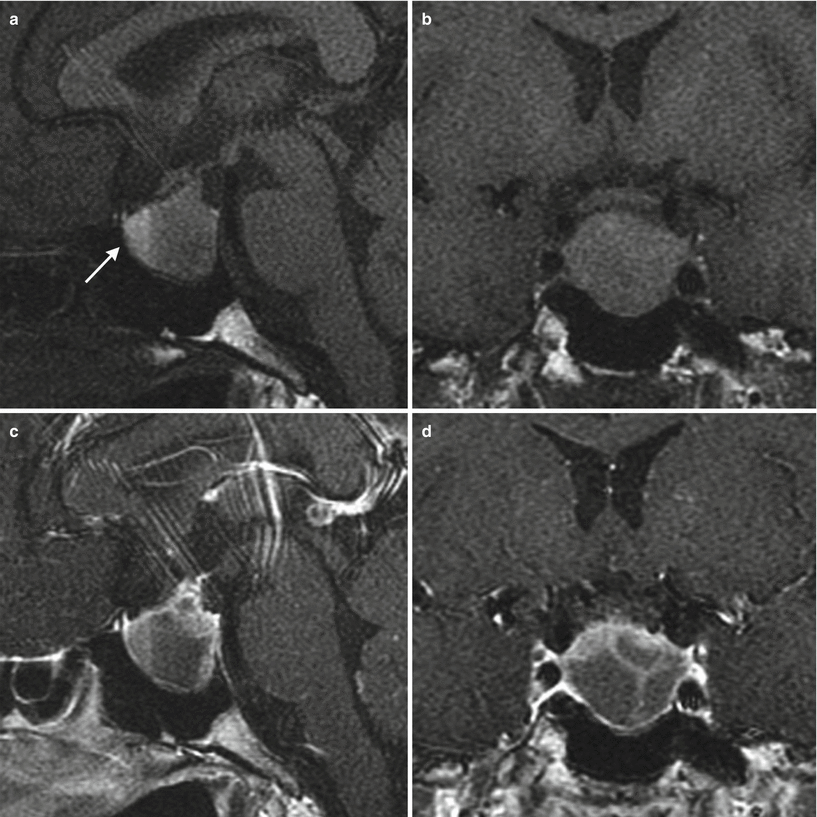
Fig. 17.1
Pituitary apoplexy. (a) Sagittal T1-weighted pre-gadolinium image. (b) Coronal T1-weighted pre-gadolinium image. (c) Sagittal T1-weighted post-gadolinium image. (d) Coronal T1-weighted post-gadolinium image. Within the sella, there is an expansile mass with heterogeneous signal intensity and areas of T1 hyperintensity that may represent proteinaceous materials or blood products. There is mucoperiosteal thickening along the roof of the right sphenoid sinus (white arrow). The pituitary stalk is elevated and deviated to the right

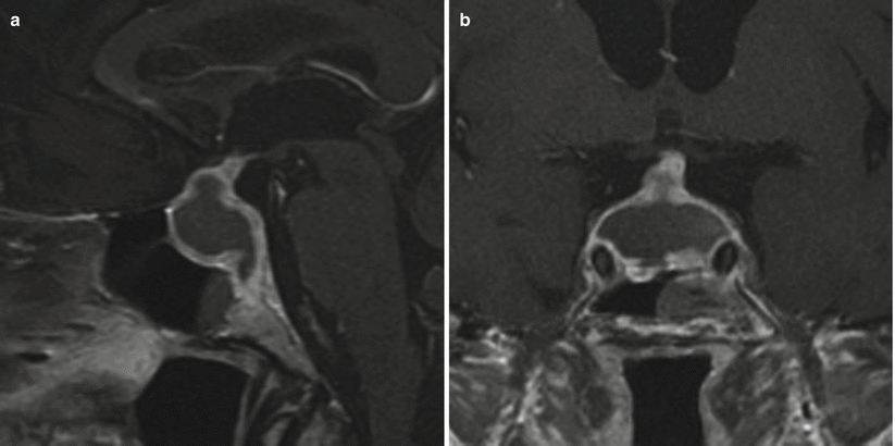
Fig. 17.2
Pituitary apoplexy. (a) Sagittal T1-weighted pre-gadolinium image. (b) Sagittal T1-weighted post-gadolinium image. (c) Coronal T1-weighted post-gadolinium image. Within the sella, there is a large sellar mass with suprasellar extension, containing a septated region of T1 hyperintensity that may represent proteinaceous materials or blood products. There is enhancing soft tissue along the periphery in the inferior and posterior aspect of the sella

Fig. 17.3
Pituitary apoplexy. (a) Sagittal T1-weighted pre-gadolinium image. (b) Coronal T1-weighted pre-gadolinium image. (c) Sagittal T1-weighted post-gadolinium image. (d) Coronal T1-weighted post-gadolinium image. Within the sella, there is an expansile mass with layering T1 hyperintensity (white arrow), which may represent proteinaceous materials or blood products. Enhancing septations are seen within the mass. There is erosion of the sellar floor

Fig. 17.4
Pituitary apoplexy. (a) Sagittal T1-weighted post-gadolinium image. (b) Coronal T1-weighted post-gadolinium image. Within the sella, there is an expansile mass with peripheral enhancement and central cyst or necrosis. Superiorly, the mass abuts the stalk without significant mass effect. There is erosion of the sellar floor. A mucous retention cyst is seen in the right sphenoid sinus

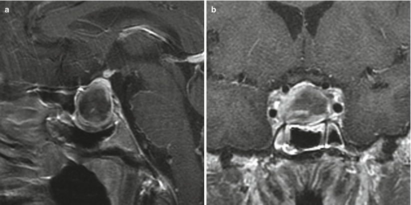
Fig. 17.5
Pituitary apoplexy. (a) Sagittal T1-weighted post-gadolinium image. (b) Coronal T1-weighted post-gadolinium image. Within the sella, there is a lobular, expansile mass with peripheral enhancement and central cyst or necrosis. The mass extends superiorly, abutting and mildly elevating the optic chiasm. There is erosion of the sellar floor and mucoperiosteal thickening along the roofs of the sphenoid sinuses


