Fig. 19.1
Pituitary carcinoma. (a) Sagittal T1-weighted pre-gadolinium image. (b) Coronal T1-weighted post-gadolinium image. (c) Sagittal T1-weighted post-gadolinium image. (d) Coronal T1-weighted post-gadolinium image. There is a large, heterogeneously enhancing sellar mass with erosion of the sella, as well as suprasellar and retroclival extensions. The mass extends anteriorly to involve the sphenoid sinus (arrow in a). There are additional parenchymal enhancing masses in the occipital lobe and corpus callosum (white arrowheads in c, d). The pituitary stalk is elevated and deviated to the right

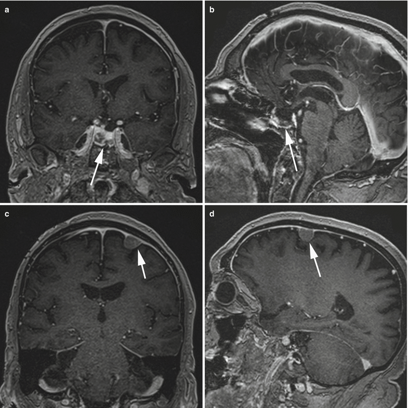
Fig. 19.2
Pituitary carcinoma. Coronal (a) and sagittal (b) T1-weighted post-gadolinium MRI in a patient with a treated atypical prolactinoma and a rising prolactin level. The sellar region (arrow) shows some residual tumor, postoperative changes, scar tissue, and radiation effects. Coronal (c) and sagittal (d) T1-weighted post-gadolinium MR images show a dura-based mass (arrow) consistent with pituitary carcinoma originating from a prolactinoma. The mass was verified histologically and subsequently resected surgically
19.2.2 Pathology
Malignant features are usually observed, with a high frequency of cellular atypia and mitotic activity [13].
Half of primary carcinoma tumors and the majority of metastases demonstrate nuclear pleomorphism and/or hyperchromasia [4].
Pituitary carcinoma has been associated with a higher mitotic index, p53 immunoreactivity, and a higher MIB-1 labeling index [4, 9, 13–16].
The mean MIB-1 index (reported as 8–11.9 % in metastatic pituitary carcinomas) is typically higher in the metastatic components [4, 13, 14].
Pituitary carcinomas have been reported to occur in almost all cell types of pituitary tumors, although they most commonly develop in ACTH adenomas and prolactinomas [1, 3].
Malignant features may be identified in atypical adenomas without the presence of metastases, but these features do not meet the qualification for the diagnosis of pituitary carcinoma [17].
19.3 Clinical Management
Treatment is frequently palliative and consists of aggressive surgical resection followed by management with chemotherapy with or without radiotherapy or radiosurgery [4].
In appropriate cases, medical therapy such as dopamine agonists and somatostatin analogs should be continued [12].
According to some recent reports, temozolomide has demonstrated some efficacy in tumor control [18–20].
Prognosis remains generally poor, however, with a mean survival time of 2–4 years; 33 % of patients do not survive 1 year beyond the time of diagnosis [4, 12].
One case report described a patient who survived 33 years with pituitary carcinoma [5].


