Fig. 9.1
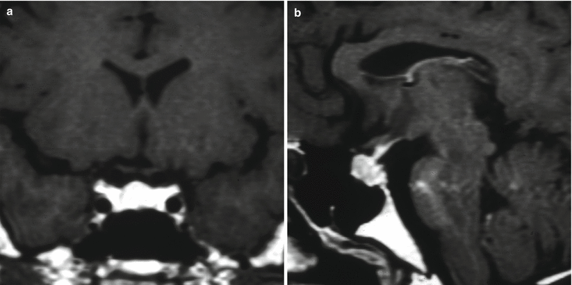
(a) Sagittal T1-weighted image post gadolinium contrast enhancement. (b) Coronal T1-weighted image post gadolinium contrast enhancement. The pituitary gland exhibits homogeneous enhancement with a convex upper margin and a craniocaudal measurement exceeding 1 cm. No focal mass or hypoenhancement is seen

Fig. 9.2
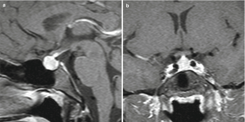
(a) Coronal T1-weighted image post gadolinium contrast enhancement. (b) Sagittal T1-weighted image post gadolinium contrast enhancement. The pituitary gland exhibits homogeneous enhancement with a convex upper margin and craniocaudal measurement exceeding 1 cm. No focal mass or hypoenhancement is seen

Fig. 9.3
(a) Sagittal T1-weighted image post gadolinium contrast enhancement. (b) Coronal T1-weighted image post gadolinium contrast enhancement. The pituitary gland exhibits homogeneous enhancement with a convex upper margin and craniocaudal measurement exceeding 1 cm. No focal mass or hypoenhancement is seen
Parasellar and infrasellar invasion are extremely rare.
9.4 Histopathology
It may be difficult to differentiate pituitary hyperplasia from an adenoma. The reticulin stain can often be used to help this differentiation [18]. In hyperplasia, the normal acinar structure of the pituitary gland is maintained but it is enlarged, whereas adenomas typically demonstrate disruption of the reticular acinar structure.
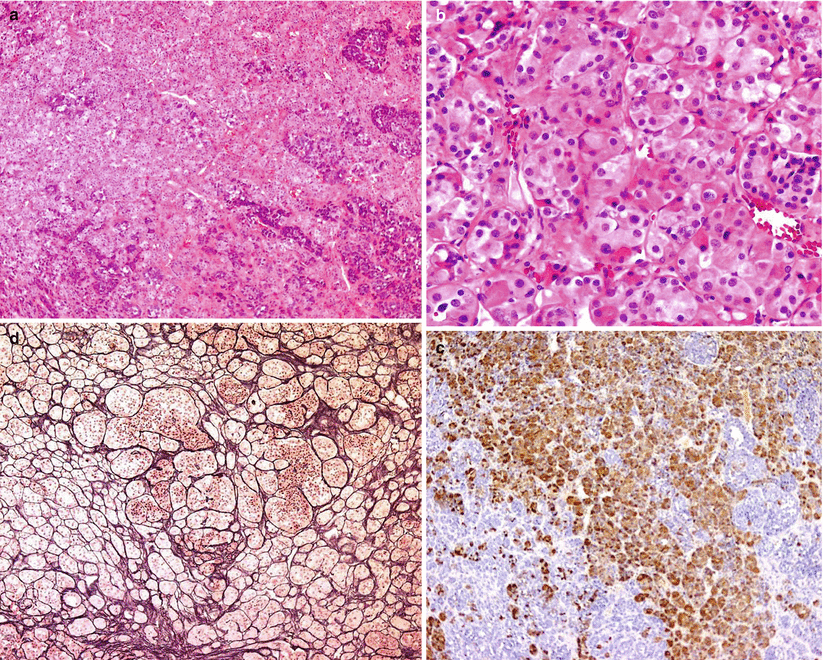
Histopathology may demonstrate diffuse or nodular hyperplasia, typically identifiable with a silver/reticulin stain [6]. In diffuse hyperplasia, an increase in the size of glandular cells and acini is seen throughout the gland, without disruption of the acinar network [18] (Fig. 9.4). Nodular hyperplasia appears as a local, nodular growth of pituitary cells, with enlargement and possible disruption of the acinar network. Thyrotroph hyperplasia is diffuse in 69 % of cases and nodular in 25 % [10]. Lactotroph hyperplasia is generally of the diffuse type [6].


Fig. 9.4
Surgical specimen from a patient with a history of hypothyroidism and a nonfunctioning adenoma found incidentally after a trauma to the head. (a, b) In addition to a null-cell adenoma (not shown), the adjacent normal gland showed expansion of the acini, which were composed mostly of acidophilic cells. (c) Reticulin stain emphasized expansion of the acini as compared with the normal pituitary pattern. (d) Immunostain for thyroid-stimulating hormone (TSH) confirmed the expansion of the thyrotroph cell population due to uncontrolled hypothyroidism
9.5 Clinical Management
When clinical and imaging characteristics lead to suspicion of pituitary hyperplasia, the underlying hormonal cause should be sought. If the inciting cause is identified and treated, pituitary hyperplasia is typically reversible. For example, thyrotroph hyperplasia is generally reversible following treatment with thyroid hormone replacement [9]. Lactotroph hyperplasia is partially reversible following pregnancy or lactation. Nulliparous women have less massive pituitary glands than multiparous women [19].
In the rare cases when mass effect results in severe headache, visual loss, or panhypopituitarism, surgical intervention for tissue diagnosis and decompression may be indicated.
References
1.
Grubb MR, Chakeres D, Malarkey WB. Patients with primary hypothyroidism presenting as prolactinomas. Am J Med. 1987;83:765–9.CrossRefPubMed
Stay updated, free articles. Join our Telegram channel

Full access? Get Clinical Tree


