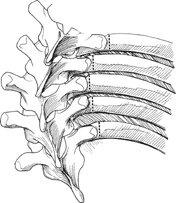
55 Bernard A. Rawlins A technique designed to provide additional mobility (release) to the spinal elements in a rigid deformity. The level of release should include the rib of the apical vertebrae and ribs caudal and cephalad, depending on the curve magnitude. Concave rib osteotomies provide additional release to the spine beyond that provided by facetectomies, anterior diskectomies, and convex rib thoracoplasty. This becomes apparent during translation maneuvers using sublaminar wires. Rigid coronal curves in adult and pediatric deformities. Severe pulmonary compromise for which surgery to both pulmonary cavities would compromise the patient’s perioperative recovery. Pulmonary function testing preoperatively is usually done in large curves and provides an assessment of pulmonary reserve. Positive pressure ventilation should be requested to evaluate air leaks following the osteotomies. Rib synostosis, particularly in a congenital curve, makes dissection difficult. Comprehensive preoperative radiographic evaluation helps avoid surprises. Always obtain a postoperative chest radiograph to search for a hemothorax and pneumothorax. If a pneumothorax is suspected either intraoperatively or postoperatively, tube thoracostomy should be performed. Standard posterior exposure to the tips of the transverse processes from the caudal to the cephalic level of the spine under consideration for fusion is performed. The sequence for a routine posterior instrumented fusion is the following: place the pedicle screws, perform the facetectomies, prepare sites and place hooks, perform laminotomies for sublaminar wire placement, perform the thoracoplasty, and then consider the concave rib osteotomies. Exposure is then carried lateral to the tips of the transverse process of the vertebrae under consideration. The dissection proceeds in a plane directly over the ribs for 2 to 3 cm. The paraspinal muscles give comparatively little resistance, given the surgeon is on the concave side of the curve. Subperiosteal dissection of the rib is performed as in a standard thoracotomy approach using the Alexander and Doyen instruments. A rib cutter is used to perform the rib osteotomy (Fig. 55.1). The area is filled with saline, and positive ventilation provided by the anesthesiologist is used to check for air leaks. A chest tube is placed immediately if an air leak is found. During the correction maneuver the rib lateral to the osteotomy should be elevated above the medial portion of the rib, transverse processes, or spinal instrumentation, as the spine is corrected in the direction of the rib osteotomy sites (Fig. 55.2).
Posterior Rib Osteotomy for Rigid Coronal Spinal Deformities
Description
Key Principles
Expectations
Indications
Contraindications
Special Considerations
Special Instructions, Position, and Anesthesia
Tips, Pearls, and Lessons Learned
Difficulties Encountered
Key Procedural Steps
Related posts:
 Wound VAC Management for Spinal or Bone Graft Wound Infections
 Reduction Techniques for Atlantoaxial Rotary Subluxation
 Posterior Spinal Anchor Strategy Placement and Rod Reduction Techniques: Posterior (Rotation vs. In-Situ Translation)
 Iliosacral Screw Fixation Techniques
 Open Lumbar Microscopic Diskectomy
 Spondylolysis Repair (Pars Interarticularis Repair)
Stay updated, free articles. Join our Telegram channel

Full access? Get Clinical Tree


