Principles of Pain Management

Chapter 44 Principles of Pain Management


Definition and Challenge


Chronic pain constitutes a major public health burden. Between 15% and 20% of the population in the United States suffers from chronic pain. The prevalence of persistent chronic pain increases with age, and between ages 35 and 54 years, the prevalence could be up to 29%. In those 55 years and older, the prevalence could be as high as 39%. Treatment of chronic headache, trigeminal neuralgia (TN), and pain conditions caused by damage or malfunction in the central and peripheral nerve systems are still a major task and challenge facing most neurologists in their daily work.


The International Association for the Study of Pain defines pain as an unpleasant sensory and emotional experience associated with actual or potential tissue damage, or described in terms of such damage, or both. Pain is classified as acute, chronic, or malignant. Acute pain is caused by injury, surgery, illness, trauma, or painful medical procedures. It generally lasts for a short period of time and usually disappears when the underlying cause has been treated or has healed. However, acute pain may lead to chronic pain problems that exist beyond an expected time for healing. Chronic pain is a persistent pain state not associated with malignancy or acute pain caused by trauma, surgery, infection, or other factors. Malignant pain is associated with carcinoma. Pain in cancer patients can be caused by disease itself, treatment, or autoimmune antibodies associated with the malignancy.


Over the last 2 decades, an increasing number of healthcare professionals outside the field of neurology are devoting more effort to the research and management of headache and other chronic pain conditions. Among them are anesthesiologists, physiatrists, and psychiatrists, as well as physicians from other specialties. Neurologists are traditionally well trained in anatomical localization and differential diagnosis of a variety of neurological disorders. In terms of pain management, neurologists are probably in a better position to identify the pain sources. However, the field of pain management has developed rapidly. Successful treatment of chronic pain conditions not only requires an accurate diagnosis, it also requires the treating physician to be familiar with new techniques available for pain management. Multidisciplinary team care is now recognized to be crucially important in the management of chronic pain, and the word multidisciplinary implies not just the involvement of physicians from different specialties, but the utilization of various treatment modalities as well. These modalities include pharmacological therapy, physical therapy and rehabilitation, psychological care, interventional pain management, alternative medicine techniques, and surgical treatment.


In this chapter, we will first outline the anatomical basis of chronic pain conditions and some recent developments in molecular pain research. The second portion of the chapter will discuss some common pain conditions seen in daily neurology practice. The last section of the chapter will illustrate recent developments in pharmacological treatment, as well as interventional pain management techniques for treating common chronic pain conditions.


Anatomy and Physiology of the Pain Pathways


Nociceptor receptors are found in skin, connective tissue, blood vessels, periosteum, and most of the visceral organs. These nociceptors are formed by peripheral endings of sensory neurons with various morphological features. Noxious stimuli are transduced into depolarizing current by specialized receptors congregated in the nociceptor terminals. Cutaneous nociceptors include: (1) high-threshold mechanical nociceptors (HTMs) associated with small-diameter myelinated axons (Aδ fibers), (2) myelinated mechanothermal nociceptors (MTs) (Aδ fibers), and (3) polymodal nociceptors associated with unmyelinated axons (C fibers). Polymodal nociceptors respond to mechanical, chemical, and thermal stimuli. The afferent fibers that convey nociceptive information are thinly myelinated Aδ fibers with conduction velocities of about 15 m/sec and unmyelinated C fibers with conduction velocities of 0.5 to 2 m/sec. Stimulation of afferent Aδ nociceptive fibers causes a sharp, well-localized pain sensation. Activation of nociceptive C fibers is associated with a dull, burning, or aching and poorly localized pain. Because pain impulses are conducted by small, slowly conducting nerve fibers, conventional nerve conduction velocity (NCV) studies that measure the speed of conduction of large myelinated fibers are not sensitive to abnormal function of small-diameter fibers. It is very common that patients with small-fiber neuropathy have normal NCV tests.


Most primary afferent fibers that innervate tissues below the level of the head have cell bodies located in the dorsal root ganglion (DRG) of the spinal nerves. Visceral nociceptive afferent fibers (Aδ, C fibers) travel with sympathetic and parasympathetic nerves whose cell bodies are also found in the DRG.


Axons of DRG neurons send the primary nociceptive afferents through the dorsal roots to the most superficial layers of the dorsal horns (Rexed laminae I and II) and to some of the deep laminae (Rexed V). The Aδ fibers conveying input from HTMs and MTs terminate primarily in laminae I and V; C fibers mainly terminate in lamina II. Neurotransmitters related to pain conduction include excitatory amino acids and neuropeptides, particularly substance P (Geracioti et al., 2006). The second-order neurons in the dorsal horn include cells that respond only to noxious stimuli (nociceptive specific neurons) and others (wide dynamic range [WDR] neurons) that respond to both nociceptive and non-nociceptive sensory stimuli.


Axons of most of the second-order sensory neurons associated with pain sensation cross in the anterior white commissure of the spinal cord and ascend as the spinothalamic tract in the opposite anterolateral quadrant. This tract is somatotopically organized, with sacral elements situated posterolaterally and cervical elements more anteromedially. In humans, most of the spinothalamic tract projects to the ventral posterolateral (VPL) nucleus of the thalamus as the neospinothalamic pathway, which is related to fast and well-localized pain sensation. Axons from the third-order sensory neurons in the VPL directly project to the primary sensory cortex. Some of the fibers in the spinothalamic tract synapse with neurons of the periaqueductal gray (PAG, spinoreticular pathway) and other brainstem nuclei. Fibers from these brainstem neurons join with fibers from the spinothalamic tract to project to the central or laminar nuclei of the thalamus and constitute the paleospinothalamic tract, which is related to slow and poorly localized pain and emotional response to pain stimulation.


Multiple areas of the cerebral cortex are involved in the processing of pain sensation and the subsequent behavioral and emotional responses. Recent functional magnetic resonance imaging (fMRI) and positron emission tomography (PET) scan studies indicate that the primary and secondary somatosensory cortex, thalamus, periaqueductal gray matter, supplemental motor, inferior prefrontal, and insular cortex are activated in response to painful stimulation. It is now believed that primary sensory cortex (SI) seems to play a role in basic pain processing, while secondary sensory cortex (SII) and insula are involved in higher functions of pain perception. Emotional aspects of pain perception are mediated by the anterior cingulate cortex and the posterior insula and parietal operculum.


Central Modulation of Nociception


Nociceptive transmissions are modulated at the spinal level by both local neuronal circuits and descending pathways originating in the brainstem through the dorsal horns and the spinothalamic projections. Intrasegmental and intersegmental projections arising from cells located in the Rexed laminae I and II modulate both presynaptic and postsynaptic elements of primary nociceptive afferent terminals in the spinal cord. Activation of non-nociceptive afferent fibers may suppress nociceptive transmission in the dorsal horn. This is the major component of circuitry models referred to as the gate control theory of pain transmission. The development and widespread use of the spinal cord stimulator is based on this theory.


Descending inhibitory systems appear to have three functionally interrelated neurotransmitter mechanisms: the opioid, the noradrenergic, and the serotonergic systems. Opioid precursors and their respective peptides (β-endorphin, methionine [met]-enkephalin, leucine [leu-] enkephalin, and dynorphin) are present in the amygdala, hypothalamus, PAG, raphe magnus, and the dorsal horn. Noradrenergic neurons project from the locus caeruleus and other noradrenergic cell groups in the medulla and pons. These projections are found in the dorsolateral funiculus. Stimulation of these areas produces analgesia, as does the administration (direct or intrathecal) of α2-receptor agonists such as clonidine (Khodayar et al., 2006). Many serotonergic neurons are found in the raphe magnus. These neurons send projections to the spinal cord via the dorsolateral funiculus. Administration of serotonin to the spinal cord produces analgesia, and pharmacological blockade or lesion of the raphe magnus can reduce the effects of morphine. The antinociceptive effects of antidepressants such as tricyclics and newer serotonin-norepinephrine reuptake inhibitors such as duloxetine and milnacipran are believed to reduce pain by increasing serotonin and norepinephrine concentrations in descending inhibitory pain pathways.


Opioid Receptors


Opioids are the core pharmacological treatment for acute pain. They act via receptors on cell membranes. Opioid receptors are coupled to G proteins and are thus able to effect protein phosphorylation via the second messenger system and change ion channel conductance. Presynaptically, activation of opioid receptors inhibits the release of neurotransmitters involved in pain, including substance P and glutamate. Postsynaptically, activation of opioid receptors inhibit neurons by opening potassium channels that hyperpolarize and inhibit the neuron.


Currently, there are five proposed classes of opioid receptors: µ, δ, κ, σ, and ε. µ Receptors are the main functional target of morphine and morphine-like drugs; they are present in large quantities in the periaqueductal gray matter in the brain and the substantia gelatinosa in the spinal cord. µ Receptors are also found in the peripheral nerves and skin. Activation of µ receptors results in analgesia, euphoria, respiratory depression, nausea, vomiting, and decreased gastrointestinal (GI) activity, as well as the physiological syndromes of tolerance and dependence. Two distinct subgroups of the µ receptors have been identified: µ1, found supraspinally, and µ2, found mainly in the spinal cord. The µ1 receptor is associated with the pain-relieving effects of opioids, whereas µ2 receptors mediate constipation and respiratory depression.


The δ receptor has similar central and peripheral distribution as the µ receptors. Studies have shown that δ-opioid agonists can provide relief of inflammatory pain and malignant bone pain. Meanwhile, peripherally restricted κ-opioid agonists have been developed to target κ-opioid receptors located on visceral and somatic afferent nerves for relief of inflammatory, visceral, and neuropathic chronic pain. The potential analgesic effects, combined with a possible lower abuse rate and fewer side effects than µ-receptor agonists, makes δ- and peripherally restricted κ-opioid receptor agonists promising targets for treating pain (Vanderah, 2010).


Central Sensitization


Central sensitization plays a major role in the development of neuropathic pain syndromes. First, postsynaptic depolarization in the spinal cord in response to afferent stimulation can induce removal of magnesium blockade in N-methyl-d-aspartate (NMDA) receptors such that glutamate now induces a depolarization upon receptor binding. This process is short lasting and is called wind up (Katz and Rothenberg, 2005). It is responsible for the temporal summation of inputs. The second set of changes is related to phosphorylation of the NMDA receptor, which is a key process for longer-lasting changes in the excitability of the dorsal horn neurons that produce central sensitization. This posttranslational modification of the NMDA receptors results in dramatic changes in excitability due to removal of the voltage-dependent magnesium block in the absence of cell depolarization and also to changes in channel kinetics, such as channel opening time. NMDA receptor activation allows calcium influx into the cell, which further augments signal transduction within the dorsal horn neurons by activating a number of intracellular signal transduction kinases. As a result, relatively brief C-fiber inputs initiate very rapid changes in membrane excitability. This manifests both as a progressive increase in excitability during the course of the stimulus (wind up) and as post-stimulus changes that may last for several hours (central sensitization).


Clinically, the real meaning of peripheral and central sensitization is the enhanced and prolonged pain perception to minor stimulations, or sometimes without peripheral stimulation. Once peripheral and central sensitizations are involved, the pain is usually more difficult to treat. It is now believed that peripheral and central sensitization may be involved in a wide variety of chronic pain conditions such as reflex sympathetic dystrophy, tension headache, carpal tunnel syndrome, pain after spinal cord injury (Carlton et al., 2009), and even in pain conditions previously thought to be mainly nociceptive in nature such as fibromyalgia, epicondylalgia, and osteoarthritis (Gwilym et al., 2009). The challenge to the clinician is that when trying to make a diagnosis for a pain patient, we should not only try to localize the pain source—as most clinicians always do—we should also factor in the role of peripheral and central sensitization and what the best treatment strategy will be in each case.


Common Pain Syndromes


Trigeminal Neuralgia


Trigeminal neuralgia (TN), or tic douloureux, is characterized by paroxysmal lancinating attacks of severe facial pain. TN has an incidence of approximately 4/100,000, with a large majority of cases occurring spontaneously. Both genders experience TN, but there is a slight female predominance, and the diagnosis is most common over the age of 50. Classic TN is characterized by abrupt onset and termination of unilateral brief electric shock-like pain. Pain is often limited to the distribution of one or two (commonly the second and third) divisions of the trigeminal nerve. Trivial stimuli including washing, shaving, smoking, talking, and/or brushing the teeth (trigger factors) can evoke the pain. Some areas in the nasolabial fold and/or chin may be particularly susceptible to stimulation (trigger areas). In individual patients, pain attacks are stereotyped, recurring with the same intensity and distribution. Most TN patients are symptom free between attacks, and clinical examination is usually normal. Attacks of TN occur in clusters, and remissions can last for months.


The cause of TN pain attacks is unknown. Compression of the trigeminal nerve by benign tumors and vascular anomalies may play a role in the development of clinical symptoms. Studies of surgical biopsy specimens from TN patients who had presumed vascular compression demonstrate evidence of inflammation, demyelination, and close apposition of axons (leading to the possibility of ephaptic transmission between fibers). The ignition hypothesis of Devor proposes that a trigeminal nerve injury induces physiological changes that lead to a population of hyperexcitable and functionally linked trigeminal primary sensory neurons. The discharge of any individual neuron in this group can quickly spread to activate the entire population, resulting in a sudden synchronous discharge and a sudden jolt of pain characteristic of a TN attack.


The diagnosis of TN is based primarily on a history of characteristic paroxysmal pain attacks. The White and Sweet criteria are still commonly used worldwide (Box 44.1). In the majority of TN patients, the clinical examination, imaging studies, and laboratory tests are unremarkable (classic TN). In a smaller group, TN is secondary to other disease processes affecting the trigeminal system (symptomatic TN). Because a significant percentage of patients have symptomatic TN resulting from other disease processes, diagnostic MRI studies should be part of the initial evaluation of any patient with TN symptoms. Special attention should be paid to MS plaques, tumor, and subtle vascular anomalies that may be the source of root compression. Recent studies found that high-resolution three-dimensional (3D) MRI reconstruction and magnetic resonance cisternography may provide alternative tools to better identify the presence of neurovascular compression and even measure the volume of neurovascular compression at the cerebellopontine angle and predict the prognosis after initial treatment (Tanaka et al., 2009).


Carbamazepine is the first choice for treatment of TN; both controlled and uncontrolled studies confirm its clinical efficacy. Carbamazepine monotherapy provides initial symptom control in as many as 80% of TN patients. Of those initially responding to the drug, approximately 75% will continue to have long-term control of pain attacks. Controlled studies demonstrate that baclofen and lamotrigine are superior to placebo for treatments of TN. In the experience of many clinicians, baclofen is just as effective as carbamazepine and often better tolerated. A recent study found that oxcarbazepine may be effective for those who were unresponsive to the treatment of carbamazepine (Gomez-Arguelles et al., 2008). Pregabalin may also be potentially effective. If a patient is not satisfied with single medication therapy, adding another oral medication may offer additional benefits. Intravenous (IV) lidocaine or phenytoin could be effective for some severe refractory cases of TN. However, these treatments carry additional risks and require close cardiovascular monitoring. Opioid analgesics have not been proven effective for TN and should be avoided.


Posterior fossa exploration and microvascular decompression (MVD) is assumed to directly treat the cause of TN. However, this is a complex and invasive therapy with a possibility of death. With the availability of other less-invasive procedures, MVD is infrequently used and is only reserved for younger and healthier patients. Several studies demonstrated trigeminal radiofrequency rhizotomy successfully controls symptoms in over 85% of TN cases. The technique is minimally invasive. To heat the gasserian ganglion, a radiofrequency needle is inserted through the foramen ovale under the guidance of fluoroscopy. The procedure can be finished in less than 30 minutes in experienced hands. A few patients experience sensory loss and dysesthesia (analgesia dolorosa) in the distribution of the damaged trigeminal fibers with this procedure. Stereotactic radiosurgery (SRS) employs computerized stereotaxic methods to concentrate ionizing radiation on the trigeminal root entry zone. Several studies have demonstrated the high clinical efficacy and relative safety of this new technique. It is currently recommended as a first-line noninvasive surgical technique in many pain centers, especially for frail or elderly patients (Zahra et al., 2009).


Low Back Pain


Low back pain (LBP) is the most common condition seen in pain clinics. Approximately 60% to 80% of the U.S. population will experience back pain some time during life. Neurologists are often consulted for the diagnosis and treatment of LBP. It is critical for clinicians to appropriately examine the patients and make a diagnosis before treatment is rendered. Common causes of LBP include muscle strain, lumbar disk herniation, lumbar radiculopathy, lumbar facet joint syndrome, sacroiliac joint syndrome, and lumbar spinal stenosis.


Patients with acute muscle strain in the low back often have histories of acute injury. Physical examination may reveal tenderness or muscle spasms. Nonsteroidal anti-inflammatory drugs (NSAIDs), muscle relaxants, massage therapy, physical therapy, or acupuncture often provide effective pain relief. However, many times muscle pain in the low back is secondary to injuries in deeper tissues, such as lumbar disk herniation or lumbar radiculopathy.


Acute lumbar disk herniation after injury may cause severe LBP. Patients often complain of severe shooting or stabbing pains in the low back, with frequent radiation pain down the dorsomedial part of the foot when the L5 nerve root is involved, or the lateral part of the foot or the small toe when the S1 nerve root is involved. The straight leg raising test is often positive. Detailed neurological examinations may find decreased sensation to pin prick in the area innervated by L5 and/or S1 nerve root(s). Patient may also have mild weakness on the tibialis anterior (L5), or peroneus longus and brevis muscles (S1). These patients usually have severe tenderness and spasm over the lumbar paraspinal muscles. Lumbosacral MRIs may reveal disk herniation at L4-5 and/or L5-S1 level(s). Electromyography (EMG)/NCV tests may not detect a lumbar radiculopathy. NSAIDs, muscle relaxants, and physical therapy may help some patients with acute disk herniation and lumbar radiculopathy. If patients fail these treatments, lumbar epidural corticosteroid injections may offer fast and effective pain relief if the nerve roots are not severely mechanically compressed. Surgery is suggested for those with severe focal weakness of relevant muscles or incontinence. Surgery may also be indicated for severe pain that lasts for more than 3 months and does not respond to aggressive pain management if disk herniation is demonstrated by MRI or computed tomography (CT) studies.


Lumbar facet joint syndrome is found in up to 35% of patients with LBP. It is frequently associated with arthritis or injuries in lumbar facet joints. Patients may complain of pain in the low back, often on one side only. Pain may radiate down the back or front of the thigh. Physical examination may find positive tenderness over the lumbar paraspinal muscles and facet joints. Back extension and lateral rotation to the side of the pain often increases the back pain. Results of a straight leg raising test should be negative. Neurological examination should be normal unless there is a coexistent lumbar radiculopathy or other neurological condition. Diagnosis of facet joint syndrome is clinical. MRI and CT reports of facet joint arthropathy do not correlate with clinical findings. Often these changes are age related. NSAIDs should be tried for patients with lumbar facet joint syndrome before they are considered for diagnostic medial branch blocks or intra-joint corticosteroid injections.


Sacroiliac (SI) joint syndrome is another major source of LBP. The patient may have pain in one side of the low back, with pain radiation down to the hip or thigh. Pain is often increased when these patients try to walk upstairs. Physical examination may find tenderness over the SI joint, and the Patrick test or single-leg standing often exacerbate SI joint pain. NSAIDs are the first-line medication for SI joint inflammation. SI joint corticosteroid injection can provide temporary pain relief. Radiofrequency lesions to denervate the SI joint have been reported effective; however, more studies are needed to confirm clinical efficacy of this treatment.


Lumbar spinal stenosis is a common age-related change. The majority of seniors older than 60 years of age have varying degrees of spinal stenosis due to disk herniation, osteophytes, or degenerative spondylolisthesis. Preexisting congenital lumbar canal stenosis predisposes to the development of this syndrome. Fortunately, fewer than 30% of those with spinal stenosis have clinical pain. Patients often have pain in the low back, with pain radiation down the back of both legs. Standing or walking may worsen pain. Patients often walk with a hunched back and sit down after walking a short distance to relieve pain (neurogenic claudication). The pain usually takes minutes to disappear, compared to seconds with vascular claudication. On physical examination, patients often have less tenderness over the lumbar spine than those with acute lumbar disk herniation. A straight leg raising test may be normal. The condition must be distinguished from vascular claudication. Patients may try NSAIDs first. Lumbar epidural corticosteroid injections may provide pain relief for this group of patient for weeks or even months. If a patient has severe pain and refuses surgery, chronic narcotic treatment often provides adequate pain control but runs a risk of the development of tolerance and addiction.


Cervicogenic Headache


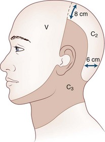
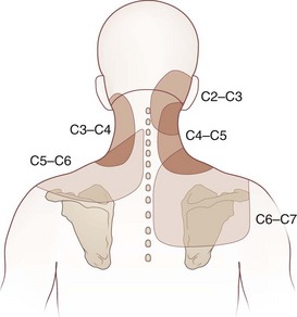
Cervicogenic headache refers to head pain originating from pathology in the neck. It is believed that pain from the C2-C3 nerve dermatome can radiate to the head and face (Fig. 44.1). An earlier study found that pain from the C2-C3 and C3-C4 cervical facet joints also can radiate to the occipital area (Fig. 44.2). The term cervicogenic headache was first introduced by Sjaastad and colleagues in 1983. However, the concept of cervicogenic headache is controversial and not well accepted by the majority of neurologists. The International Headache Society (2004) published its first diagnostic criteria in 1998 and revised it 2004. Patients with cervicogenic headache often have histories of head and neck trauma. Pain may be unilateral or bilateral. Pain is frequently localized to the occipital area, but it may also be referred to the frontal, temporal, or orbital regions. Headaches may be triggered by neck movement or sustained neck postures. This headache is constant with episodic throbbing attacks like a migraine. Patients may have other symptoms mimicking a migraine, such as nausea, vomiting, photophobia, phonophobia, and blurred vision. Owing to significant overlap of the symptoms of cervicogenic headache and migraine without aura, cervicogenic headache is often misdiagnosed as migraine. Clinicians should always consider cervicogenic headache in the differential diagnoses when evaluating a headache patient. History of head/neck injury and detailed examination of the occipital and upper cervical area should be part of the evaluation for headache. Patients with cervicogenic headache may have tenderness over the greater or lesser occipital nerve, cervical facet joints, and muscles in the upper or middle cervical region. Cervicogenic headache does not respond well to migraine medications. Treatment should be focused on removal of the pain source from the occipital-cervical junction. Initial therapy is directed to physical therapy modalities and NSAIDs. Interventional treatment such as greater occipital nerve block, cervical facet joint block, deep cervical plexus block, and botulinum toxin injections may provide effective pain relief (Zhou et al., 2010).


Complex Regional Pain Syndrome


Terminology describing the complex regional pain syndromes has evolved over the last century. The term causalgia was first coined by Weir Mitchell in the 1870s for severe progressive distal limb pain with major nerve injury. In 1946, Evans introduced the term reflex sympathetic dystrophy (RSD); it was later defined by the International Association for Study of Pain (IASP) as “continuous pain in a portion of an extremity after trauma, which may include fracture but does not involve a major nerve, associated with sympathetic hyperactivity.” In 1994, the IASP introduced the term complex regional pain syndrome (CRPS), describing a painful condition that includes regional pain, sensory changes (e.g., allodynia), abnormalities of temperature, abnormal sudomotor activity, edema, and abnormal skin color changes that occur after an initiating noxious event such as trauma. Two types of CRPS have been recognized: CRPS I corresponds to RSD, in which no definable nerve lesion is found. CRPS II refers to the cases with a definable nerve lesion and corresponds to the earlier term of causalgia.


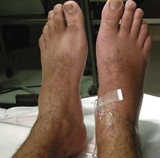
The mean age of CRPS patients ranges from 36 to 46 years, with women predominating (60%-81%). It is caused typically by an injury such as a fracture (16%-46%), strain or sprain (10%-29%), post surgery (3%-24%), and contusion or crush injury (8%-18%). Clinical features of CRPS often include pain, edema, autonomic dysfunction such as change in temperature or color in the involved limbs, motor dysfunction, and psychological abnormalities such as depression (Fig. 44.3). Schwartzman and Maleki reported the pattern of spreading of CRPS in three stages. In the early stage, CRPS often involves only one limb with pain, minor edema, and increased skin temperature. CRPS may spread from one limb to the others. In the later stage, CRPS could involve the full body and the four extremities with severe pain, edema, cold and cyanotic limbs, joint contracture, and atrophy of muscles and bones.


Excruciating pain is the cardinal feature of CRPS. Pain is often described as burning, aching, pricking, or shooting. Severity of pain is not proportional to the initial injury, and pain is not limited to the area of the injury or a specific nerve distribution. Patients may feel severe pain to minor pain stimulation such as a safety-pin prick (hyperalgesia). A light touch to skin (innocuous stimulation) may cause severe long-lasting pain (mechanical allodynia). A cooling stimulus such as a drop of alcohol may be perceived as painful (thermal allodynia). Decreased temperature and pinprick sensations in the affected limb are common.


Edema of the affected limb is present in the majority of patients. It could be very mild in the early stage of CRPS, mimicking mild cellulitis. However, in the late stage, edema may be so severe that a Doppler test is needed to rule out the possibility of deep vein thrombosis.


Autonomic dysfunction may manifest as changes of skin color and temperature, as well as sweating abnormalities. The affected area may be reddish at one time and then become blue, purple, or pale over a course of minutes to hours. Livedo reticularis is common in CRPS. Livedo is a descriptive term used to describe the red, non-blanchable (i.e., does not turn white when pressed) network pattern (reticulated) in the skin. About 60% of patients may report excessive sweating in the affected limbs. Temperature asymmetry between the affected and unaffected sides may exceed 1°C.


Motor dysfunctions in CRPS include mild weakness, decreased range of motion, tremor, dystonia, and myoclonus. Dystrophic manifestations are seen in the form of increased or decreased nail and hair growth in the affected extremity, hyperkeratosis or thin glossy skin, and osteoporosis of the underlying bones.


Diagnosis of CRPS is clinical. According to IASP, if a patient has the above-mentioned features, a diagnosis of CRPS may be made if other clinical conditions such as infection or DVT are ruled out. EMG/NCV tests are not sensitive to CRPS and frequently cause severe pain to patients. A triple-phase bone scan may reveal abnormal absorption in the affected limbs (increased or decreased), though it is not a primary diagnostic procedure for CRPS.


The pathophysiology of CRPS is not completely understood. Multiple mechanisms are considered in the generation and maintenance of CRPS. Increased systemic calcitonin gene-related peptide (CGRP) levels may contribute to neurogenic inflammation, edema, vasodilatation, and increased sweating. Elevated neuropeptide concentrations may lead to pain and hyperalgesia. Immunological mechanisms (e.g., altered expressions of human leukocyte antigen [HLA], substance P, cytokines, and interleukins) are believed to contribute to the pathogenesis of clinical symptoms such as edema. Up-regulation of adrenergic receptors and functional coupling between sympathetic efferent and sensory afferent fibers may provide the basis of the sympathetic nervous system abnormalities in the pathogenesis of CRPS. The central mechanisms in CRPS may include central sensitization in the spinal cord, brainstem, or thalamus, cortical reorganization in the primary somatosensory cortex, and disinhibition of the motor cortex.


The goals of treatment for CRPS are pain relief, functional recovery, and psychological improvement. However, treatment of CRPS remains a challenge. There is little if any evidence for the efficacy of any treatment modality. In the early stages of CRPS treatment, occupational and physical therapies are often used. Occupational and physical therapies are supported by anecdotal data and have not been validated by randomized prospective trials.


Patients diagnosed with CRPS for over 2 months should also undergo a psychological evaluation—which includes psychometric testing—to identify and treat psychological disorders such as anxiety, depression, or personality disorders. Counseling, behavioral modification, biofeedback, relaxation therapy, group therapy, and self-hypnosis should be considered. The goal of psychotherapy is to improve patient motivation and coping skills.


Tricyclic antidepressants, antiepileptics, and narcotics such as methadone are commonly used empirically for CRPS, even though clinical controlled studies have not proven their efficacy. A recent review article summarized the evidence derived from randomized controlled trials pertaining to the treatment of CRPS. The review reported clinical improvement with dimethyl sulfoxide, steroids, epidural clonidine, and intrathecal baclofen. Only bisphosphonates appear to offer clear benefits for patients with CRPS (Tran de et al., 2010). NMDA receptor modulation is a major interest of current research. It has been reported that subanesthetic infusions of ketamine might offer a promising therapeutic option in the treatment of appropriately selected patients with intractable CRPS (Schwartzman et al., 2009). A recent preliminary study reported that IV immunoglobulin treatment could potentially decrease pain in CRPS patients (Goebel et al., 2010). However, more studies are needed to further establish the safety and efficacy of these novel approaches.


Minimally invasive techniques have been used extensively for the treatment of CRPS. Techniques include sympathetic block, intravenous regional block (IVRB), somatic nerve block, epidural drug administration, intrathecal drug delivery, and neurostimulation. Stellate ganglion blocks in early-stage CRPS may significantly decrease pain and hasten clinical recovery. It may also prevent the recurrence of CRPS after reoperation of the affected extremity. In a double-blind study, IVRB with bretylium provided significantly longer analgesia than lidocaine. Good pain relief is reported with the use of epidural delivery of clonidine and ketamine and also with intrathecal baclofen and morphine. An early study with 2-year follow-up reported that spinal cord stimulation (SCS) results in a long-term pain reduction and improvement in health-related quality of life. However, a more recent randomized study with 5-year follow-up found no extra benefit in terms of pain relief for those with a combination of SCS and physical therapy, compared to those with physical therapy alone (Kemler et al., 2008). The author shares the same experience and opinion with the cited report. It seems that most RSD patients feel better immediately after the SCS implantation. However, the SCS itself may have difficulty stopping the spread of RSD, and once RSD spreads out of the area initially covered by the SCS, the pain is no longer “under control.”


Post-Stroke Pain Syndrome


Lesions at any level of the neuroaxis (generally affecting spinothalamocortical afferent sensory pathways) including the medulla, pons, midbrain, thalamus, subcortical white matter, and the cortex may produce central post-stroke pain syndrome (PSP). However, the thalamus and brainstem are common sites for PSP; 8% to 16% of thalamic stroke may lead to chronic pain. The frequency of pain after a geniculothalamic artery stroke is even higher (13%-59%).


The pathogenesis of PSP is not yet known. However, it has been suggested that hyperexcitation in the damaged sensory pathways, damage to the central inhibitory pathways, or a combination of the two may be responsible for the onset of PSP. Pain is the cardinal symptom and is described as spontaneous, severe, paroxysmal, and burning. Patients with thalamic pain syndrome also have hyperalgesia and allodynia in the affected limbs. Right-sided lesions predominate among reported cases of the thalamic pain syndrome.


Patients reporting pain due to brainstem infarction usually have involvement of pontine or medullary structures. Patients with midbrain infarction seldom complain of pain. Transitory eye and nose pain may be an initial symptom of pontine infarction. About 25% patients with dorsolateral medullary infarction develop ipsilateral facial pain, especially when the lesion involves the spinal trigeminal tract. Facial allodynia is also common. Some patients may experience pain in the contralateral limbs and trunk.


Treatment of central post-stroke pain remains a challenge. Tricyclic antidepressants are still a choice of treatment. Gabapentin and lamotrigine have been used to treat central post-stroke pain syndrome in open-labeled studies. Selective posterior rhizotomy has been reported to decrease painful spasticity in the lower limbs of hemiplegic patients after a stroke. It has been reported that chronic motor cortex stimulation therapy provides pain relief for some post-stroke patients (Brown and Pilitsis, 2006). Stereotactic radiosurgery of the pituitary and deep brain stimulation (DBS) have been used to treat PSP syndrome with some success (Pickering et al., 2009).


Some 40% to 60% of patients develop shoulder pain after a stroke. The mechanism of shoulder pain is not clear, but a strong association exists between pain and an abnormal shoulder joint examination, ipsilateral sensory abnormalities, and arm weakness. These patients usually have significant tenderness over the shoulder joint. It is postulated that the pain is due to inflammation in the joint secondary to immobilization and joint contracture ( frozen shoulder syndrome). The majority of shoulder pain may be resolved or improved for 6 months following a stroke with intensive physical/occupational therapy. Antiinflammatory medications may be used. Suprascapular nerve or brachial plexus block can provide temporary pain relief to prepare for physical therapy. Proper positioning of the shoulder, range-of-motion activities, and avoidance of immobilization may further help prevent or alleviate shoulder pain.

< div class='tao-gold-member'>

Stay updated, free articles. Join our Telegram channel

Jun 19, 2016 | Posted by in NEUROLOGY | Comments Off on Principles of Pain Management

Full access? Get Clinical Tree

Get Clinical Tree app for offline access