Proximal, Distal, and Generalized Weakness

Chapter 25 Proximal, Distal, and Generalized Weakness


Muscle weakness may be due to disorders of the central nervous system (CNS) or peripheral nervous system (PNS). The PNS includes the primary sensory neurons in the dorsal root ganglia, nerve roots, peripheral nerves, neuromuscular junctions, and muscles. Although not strictly peripheral, the primary motor neurons (anterior horn cells) in the brainstem and spinal cord are also conventionally included as part of the PNS. The neurological examination allows separation of the causes of weakness arising at these different locations. If the pattern of weakness is characteristic of upper motor neuron (UMN) dysfunction (i.e., weakness of upper-limb extensors and lower-limb flexors) together with hyperreflexia and extensor plantar responses, the weakness clearly is of CNS origin. Weakness with sensory loss may occur in both CNS disorders and disorders of the nerve roots and peripheral nerves. Weakness without sensory loss may also occur from CNS disorders, but in the PNS, this pattern of weakness occurs in disorders of the anterior horn cell, neuromuscular junction, or muscle. Rarely in the PNS, peripheral motor fibers will be the site of pathology (e.g., as occurs in multifocal motor neuropathy with conduction block). Although fatigue often accompanies most disorders of weakness, marked fatigue, especially when involving the extraocular, bulbar, and proximal upper limb muscles, often indicates a disorder of the neuromuscular junction.


The motor unit is the primary building block of the PNS and includes the anterior horn cell, its motor nerve, terminal nerve fibers, and all their accompanying neuromuscular junctions and muscle fibers. This chapter concentrates on disorders of the motor unit and disorders that may also involve the peripheral sensory nerves. The pattern of weakness often localizes the pathological process to the primary neurons, nerve roots, peripheral nerves, neuromuscular junctions, or muscles. Muscle weakness changes functional abilities that are more or less specific to the muscle groups affected. Recognizable patterns of symptoms and signs often allow a reasonable estimation of the anatomical involvement. Identifying these patterns is the first step in the differential diagnosis of weakness, because certain disorders affect specific muscle groups. This chapter begins with a review of the symptoms and signs of muscular weakness with respect to the muscle groups affected. A discussion follows of the bedside examinations, functional examinations, and laboratory tests often used in evaluating patients with muscle weakness. The chapter concludes with an approach to the differential diagnosis of muscle weakness based on which muscle groups are weak, whether the muscle weakness is constant or fluctuating, and whether the disorder is genetic or acquired.


Clinical Presentation by Affected Region


General Considerations


As muscles begin to weaken, the associated clinical features depend more on which muscles are involved than on the cause of involvement. A complicating factor in evaluating weakness is the patient’s interpretation of the term weak. Although physicians use this term to denote a loss of muscle power, patients tend to apply it more loosely in describing their symptoms. Even more confusing, many people use the words numb and weak interchangeably, so the clinician should not accept a complaint of weakness at face value; the patient should be questioned further until it is clear that weakness means loss of muscle strength.


If the patient has no objective weakness when examined, the clinician must rely on the history. In patients with weak muscles, a fairly stereotypical set of symptoms emerges according to which muscle groups are weak (discussed later in this section). The patient whose weakness is caused by depression or malingering has vague symptoms, avoids answering leading questions, and the stereotypical symptoms of weakness are seldom volunteered. Instead, these patients make such statements as “I have no strength to do the housework,” “I just can’t do (the task),” or “I can’t climb the stairs because I get so tired and have to rest.” When pressed regarding these symptoms, it soon becomes apparent that specific details are lacking. Patients who cannot get out of a low chair because of real weakness explain exactly how they have to maneuver themselves into an upright position (e.g., pushing on the chair arms, leaning forward in the seat, and bracing their hands against the furniture). The examiner should avoid providing patients with clinical details they appear to be searching for. Asking whether pushing on the arms of the chair is required to stand up provides the patient with key information that may later be used in response to the questions of baffled successive examiners. In addition, it often is difficult to differentiate true muscle weakness from apparent weakness that accompanies tendon or joint contractures or is secondary to pain. For example, patients with primary orthopedic conditions often complain of weakness. In these patients, however, pain with passive or active motion often is a prominent part of the symptoms.


In evaluating weakness, the first key task is to discern which muscle groups are affected. In this regard, it is helpful to consider the clinical presentation with involvement of specific body regions: ocular; facial and bulbar; neck, diaphragm, and axial; proximal upper extremity; distal upper extremity; proximal lower extremity; and distal lower extremity.


Facial and Bulbar Muscles


Patients experience facial weakness as a feeling of stiffness or sometimes as a twisting or altered perception in the face (note that patients often use the word numbness in describing facial weakness). Drinking through a straw, whistling, and blowing up balloons all are particularly difficult tasks for these patients and may be sensitive tests for facial weakness, particularly when such weakness dates from childhood. Acquaintances may notice that the patient’s expression is somehow changed. A pleasant smile may turn into a snarl because of weakness of the levator anguli oris muscles. In lower facial weakness, patients may have difficulty with drooling and retaining their saliva, often requiring them to carry a tissue in the hand—the so-called napkin sign—which often accompanies bulbar involvement in amyotrophic lateral sclerosis (ALS). A common observation in mild long-standing facial weakness, as with facioscapulohumeral (FSH) muscular dystrophy, is a tendency for the patient to sleep with the eyes open from weakness of the orbicularis oculi. Weakness of masticatory muscles may result in difficulty chewing, sometimes with a sensation of fatigue and discomfort, as may occur with myasthenia gravis (MG). Pharyngeal, palatal, and tongue weakness disturbs speech and swallowing. A flaccid palate is associated with nasal regurgitation, choking spells, and aspiration of liquids. Speech may become slurred or acquire a nasal or hoarse quality. In contrast with central lesions, no problem with fluency or language function is observed.


Neck, Diaphragm, and Axial Muscles


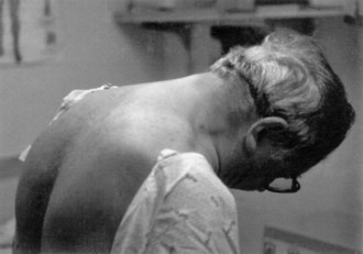
Neck muscle weakness becomes apparent when the patient must stabilize the head. Riding as a passenger in a car that brakes or accelerates, particularly in emergencies, may be disconcerting for the patient with neck weakness, because the head rocks forward or backward. Similarly, when the patient is stooping or bending forward, weakness of the posterior neck muscles may cause the chin to fall on the chest. A patient with neck-flexion weakness often notices difficulty lifting the head off the pillow in the morning. As neck weakness progresses, patients may develop the dropped head syndrome, in which they no longer can extend the neck, and the chin rests against the chest (Fig. 25.1). This posture leads to several secondary difficulties, especially with vision and swallowing.


Shortness of breath often develops when diaphragm muscles weaken, especially when individuals lie flat or must exert themselves. These symptoms can be mistakenly attributed to lung or heart disease. Severe diaphragmatic weakness leads to hypoventilation and carbon dioxide retention. This may first be manifested as morning headaches or vivid nightmares. Later, hypercapnia results in sedation and a depressed mental state. Rarely, axial and trunk muscles can be involved early in the course of a neuromuscular disorder. Weakness of the abdominal muscles may make sit-ups impossible. Focal weakness of the lower abdominal muscles results in an obvious protuberance that superficially mimics an abdominal hernia. Patients with weakness of the paraspinal muscles are unable to maintain a straight posture when sitting or standing, although they can do so when lying on the bed (so-called bent spine syndrome).


Bedside Examination of the Weak Patient


The neurological examination of patients with muscle weakness is the same as that used for patients with other neurological problems. Special attention to the observational and functional components of the evaluation, however, is particularly rewarding in the patient with weakness.


Observation


It is useful to spend a few moments observing the patient and noting natural posture and motion. When patients, particularly children, are aware of the examination, they often concentrate on performing as normally as possible. When unaware of scrutiny, their posture and movements may be more natural. At one time or another, we have heard the parent’s exasperated cry, “He never does it that way at home.” For example, ptosis may be obvious on inspection of the head and neck. The more severe the ptosis, the greater the patient’s tendency to throw the head backward. The eyebrows are elevated and the forehead wrinkled in an attempt to raise the upper lids. This sometimes is so successful that ptosis is apparent only when the examiner smoothes out the wrinkled forehead and allows the eyebrows to assume a more normal position. Psychogenic ptosis is easy to detect: the lower lid elevates with contraction of both parts of the orbicularis oculi muscles (i.e., blepharospasm) to accompany the lowered upper lid.


Weakness of the facial muscles present since childhood may give a smooth, unlined appearance to the adult face. In addition, facial expression diminishes or changes. A smile may become a grimace or a snarl, with eversion of the upper lip. The normal blink may slow, or eyelid closure may be incomplete so that the sclera is always visible. The normal preservation of the arch of the upper lip may be lost, and the mouth may assume either a tented or a straight-line configuration. Actual wasting of the facial muscles is difficult to see, but temporalis and masseter atrophy produce a characteristic scalloped appearance above and below the cheekbone. Because rearranging the hair style may cover the wasting, the examiner should make a conscious effort to check the upper portion of the patient’s face. The tongue is inspected for atrophy and fasciculations. Inspecting the tongue at rest with the mouth open, looking for the random irregular twitching movements of fasciculations, is the best method. When the tongue is fully protruded, many patients have some normal quivering movements that can easily be mistaken for fasciculations. It is wise to diagnose fasciculations of the tongue only when there is associated atrophy.


Facial weakness causes the normal labial sounds (that of p and b) to be softened. The examiner with a practiced ear can detect other alterations of speech. Lower motor neuron (LMN) involvement of the palate and tongue gives the speech a hollow, nasal, echoing timbre, whereas UMN dysfunction causes the speech to be monotonous, forced, and strained. Laryngeal weakness also may be noticed in speech when the voice becomes harsh or brassy, often associated with loss of the glottal stop (the small sound made by the larynx closing, as at the start of a cough).


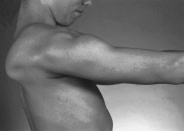
Weakness of the shoulder muscles causes a characteristic change in posture. Normally the shoulders brace back by means of the tone of the muscles, so the hands are positioned with the thumbs forward when the arms are by the side. As the shoulder muscles lose their tone, the point of the shoulder rotates forward. This forward rotation of the shoulder is associated with a rotation of the arm, so that the backs of the hands now are forward facing. Additionally, the loss of tone causes a rather loose swinging movement of the arms in normal walking. When shoulder weakness is severe, the patient may fling the arms by using a movement of the trunk, rather than lifting the arms in the normal fashion. In the most extreme example, the only way the patient can get the hand above the head on a wall is to use a truncal movement to throw the whole arm upward and forward so the hand rests on the wall, and then to creep the hand up the wall using finger movements. Atrophy of the pectoral muscles leads to the development of a horizontal or upward sloping of the anterior axillary fold. This is especially the case in facioscapulohumeral (FSH) muscular dystrophy. The examiner may observe winging of the scapula, a characteristic finding in weakness of muscles that normally fix the scapula to the thorax (i.e., the serratus anterior, rhomboid, or trapezius). As these muscles become weak, any attempted movement of the arm causes the scapula to rise off the back of the rib cage and protrude like a small wing. The arm and shoulder act as a crane—the boom of the crane is the arm, and the base is the scapula. Obviously, if the base is not fixed, any attempt to use the crane results in the whole structure’s falling over. This is the operative mechanism with attempts to elevate the arm; the scapula simply pops off the back of the chest wall in a characteristic fashion. In the most common type of winging, the entire medial border of the scapula protrudes backward. In some diseases, particularly FSH muscular dystrophy, the inferomedial angle juts out first, and the entire scapula rotates and rides up over the back. This often is associated with a trapezius hump, in which the middle part of the trapezius muscle in the web of the neck mounds over the upper border of the scapula (Fig. 25.2). Note that when examining a slender person or a child, in whom prominent shoulder blades are common, the shoulder configuration returns to normal with forcible use of the arm, as in a push-up.


Muscle Bulk and Deformities


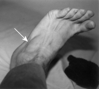
Assessment of muscle bulk looking for atrophy and hypertrophy is an important part of the neuromuscular examination. Prominent muscle wasting usually accompanies neurogenic disorders associated with axonal loss. However, severe wasting also occurs in chronic myopathic conditions. Wasting is best appreciated in the distal hand and foot muscles and around bony prominences. In the arm, wasting of the intrinsic hand muscles produces a characteristic hand posture in which the thumb rotates outward so that it lies in the same plane as that of the fingers (the simian hand), and the interphalangeal joints flex slightly with slight extension of metacarpophalangeal joints (the claw hand). Wasting of the small muscles leaves the bones easily visible through the skin, resulting in the characteristic guttered appearance of the back of the hand. In the foot, one of the easier muscles to inspect is the extensor digitorum brevis, a small muscle on the lateral dorsum of the foot that helps dorsiflex the toes (Fig. 25.3). It often wastes early in neuropathies and anterior horn cell disorders. In myopathic conditions in which proximal muscles are affected more than distal muscles, the extensor digitorum brevis may actually hypertrophy to try to compensate for weakness of the long toe dorsiflexors above it.


Muscle mass of the leg is so variable among individuals that it is sometimes difficult to decide whether wasting of the muscles has occurred. Any marked asymmetry indicates an abnormality, but distinguishing a slender thigh from quadriceps muscle atrophy often is difficult. One way to try to distinguish these conditions is to ask the patient to tighten the knee as firmly as possible. The firm medial and lateral bellies of the normal quadriceps that bunch up in the distal part of the thigh just above the knee fail to appear in the wasted muscle. The same technique can be used to evaluate anterior tibial wasting. In a severely wasted muscle, a groove on the lateral side of the tibia (which normally is filled by the anterior tibial muscles) is apparent. A moderate degree of wasting is difficult to distinguish from thinness of the leg, but if the patient dorsiflexes the foot, the wasted muscle fails to develop the prominent belly seen in a normal muscle.


Abnormal muscle hypertrophy is uncommon but may be a key finding when present. Beyond the expected increase in muscle bulk that accompanies exercise, generalized muscle hypertrophy is a feature of myotonia congenita and paramyotonia congenita, giving the appearance of the extreme development typically seen in weight lifters. Hypertrophy is a common finding in the rare syndrome of acquired neuromyotonia, in which the continuous discharge of motor axons results in the muscle effectively exercising itself. Exceptionally, hypertrophy occurs in some chronic denervating disorders, especially in the posterior calf muscle in S1 radiculopathies. Electromyography (EMG) in affected patients often reveals spontaneous discharges in these muscles (usually complex repetitive discharges) consequent to chronic denervation. By contrast, conditions exist in which muscle hypertrophy is not from true muscle enlargement but from infiltration of fat, connective tissue, and other material (i.e., pseudohypertrophy). Pseudohypertrophy occurs in calf muscles of patients with Duchenne and Becker muscular dystrophy, as well as in patients with limb-girdle muscular dystrophy, spinal muscular atrophy (SMA), and some glycogen storage disorders. Similarly, pseudohypertrophy occurs rarely in sarcoidosis, cysticercosis, amyloidosis, hypothyroid myopathy, and focal myositis. Palpable masses in muscles occur with muscle tumors, ruptured tendons, or muscle hernias.


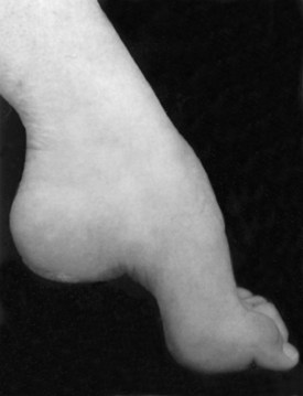
Several bony deformities often provide important clues to the presence of neuromuscular conditions. Proximal and axial muscle weakness often leads to scoliosis. Intrinsic foot muscle weakness present from childhood often leads to the characteristic foot deformity of pes cavus, in which the foot is foreshortened with high arches and hammer toes (Fig. 25.4). Pes cavus is a sign that weakness has been present at least since early childhood and implies a genetic disorder in most patients. Likewise, a high-arched palate often develops from chronic neuromuscular weakness present from childhood.


Muscle Palpation, Percussion, and Range of Motion


Palpation and percussion of muscle provide additional information. Fibrotic muscle may feel rubbery and hard, whereas denervated muscle may separate into discrete strands that roll under the fingers. Muscle in inflammatory myopathies or rheumatological conditions may be tender to palpation, but severe muscle pain on palpation is unusual. An exception to this rule is in the patient experiencing an acute phase of viral myositis or rhabdomyolysis, whose muscles may be very sensitive to either movement or touch. Percussion of muscle may produce the phenomenon of myotonia, in which a localized contraction of the muscle persists for several seconds after percussion. Percussing the thenar eminence and watching for a delayed relaxation of the thumb abductors will best show this phenomenon. This defining characteristic of myotonic dystrophy and myotonia congenita is distinguishable from myoedema, which occasionally occurs in patients with thyroid disorders and other metabolic problems. In myoedema, the development of a dimple in the muscle, which then mounds to form a small hillock, follows the percussion.


In addition to its diagnostic value, the presence of muscle contracture across a joint may cause disability, even in the absence of weakness. Thus, an evaluation of range of motion at major joints is an important part of the clinical examination. A standard examination includes evaluation for contractures at the fingers, elbows, wrists, hips, knees, and ankles. At the hips, both flexion and iliotibial band contractures should be looked for.


Muscle Tone


The physiological origin of muscle tone is complex and outside the scope of this chapter. In examining the weak patient, however, muscle tone offers valuable information regarding the origins of the weakness. Variations from a normal muscle tone result in increased tone (hypertonicity) or decreased tone (hypotonicity). Increased tone results from the loss of CNS influences on the tonic contraction of muscle. Decreased tone usually implicates a problem with the proprioceptive or peripheral motor innervation of a muscle but also may result from an acute spinal cord or cerebral lesions. Patients usually do not complain directly of increased or decreased tone; for example, the spastic patient may complain of heaviness, stiffness, or slowness of movement.


Several methods are used to examine tone. First is the spontaneous posture of the extremities. With spasticity, the upper limbs often are in a fixed flexed posture, and affected muscles are firm to palpation. The examiner should attempt to relax the patient to allow free passive movement; helpful instructions may include statements such as “Don’t try to help me do the work.” Normally, resistance is the same throughout the range of motion and does not change with changes in the velocity of the movement. In a patient with spasticity, rapid passive displacement of the extremity results in increased resistance followed by relaxation (clasp-knife phenomenon). Resistance varies with the speed and direction of passive motion. Examination of tone in the legs should include supine examination, because with the patient in this position, the examiner easily accomplishes hip and knee flexion. In spasticity, the heel elevates off the examination table, while normally the heel remains in contact with the table. Hypotonia is the loss of normal tone and is felt as increased ease of passive movements during these maneuvers, or floppiness. In patients with severe hypotonia, the joints may be passively hyperextended.


Strength


Evaluation of individual muscle strength is an important part of the clinical examination. Many methods are available. Fixed myometry has become popular within the research community. This method uses a strain gauge attached to a rigid supporting structure, often integrated into the examining couch on which the patient lies. The patient then uses maximum voluntary contraction, quantitated in newtons (N). The merits of this method are debatable, and for the average clinician, the equipment expense is prohibitive.


In an office situation and in many clinical drug trials, manual muscle testing gives perfectly adequate results and is preferable to fixed myometry in young children. The basis is the Medical Research Council grading system, with some modification (Table 25.1). This method is adequate for use in an office situation, particularly if supplemented by the functional evaluation. A scale of 0 to 5 is used, in which 5 indicates normal strength. A grade of 5 indicates that the examiner is certain a muscle is normal and never used to compensate for slightly weak muscles. Muscles that can move the joint against resistance may vary quite widely in strength; grades of 4+, 4, and 4− often are used to indicate differences, particularly between one side of the body and the other. Grade 4 represents a wide range of strength, from slight weakness to moderate weakness, which is a disadvantage. For this reason, the scale has been more useful in following the average strength of many muscles during the course of a disease, rather than the course of a single muscle. Averaging many muscle scores smoothes out the stepwise progression noted in a single muscle. This may demonstrate a steadily progressive decline. A grade of 3+ is assigned when the muscle can move the joint against gravity and can exert a tiny amount of resistance but then collapses under the pressure of the examiner’s hand. It does not denote the phenomenon of sudden give-way, which occurs in conversion disorders and in patients limited by pain. Grade 3 indicates that the muscle can move the joint throughout its full range against gravity, but not against any added resistance. Sometimes, particularly in muscles acting across large joints such as the knee, the muscle is capable of moving the limb partially against gravity but not through the full range of movement. A muscle that cannot extend the knee horizontally when the patient is in a sitting position but can extend the knee to within 30 to 40 degrees of horizontal is graded 3−. Grades 2, 1, and 0 are as defined in Table 25.1.


Table 25.1 The Medical Research Council Scale for Grading Muscle Strength


Grade Description
0 No contraction
1 Flicker or trace of contraction
2 Active movement with gravity eliminated
3 Active movement against gravity
4 Active movement against gravity and resistance
5 Normal power

Although it is commendable and sometimes essential to examine each muscle separately, most clinicians test muscle groups rather than individual muscles. In our clinic, we test neck flexion, neck extension, shoulder abduction, internal rotation, external rotation, elbow flexion and extension, wrist flexion and extension, finger abduction and adduction, thumb abduction, hip flexion and extension, knee flexion and extension, ankle dorsiflexion and plantar flexion, and dorsiflexion of the great toe.

< div class='tao-gold-member'>

Stay updated, free articles. Join our Telegram channel

Jun 19, 2016 | Posted by in NEUROLOGY | Comments Off on Proximal, Distal, and Generalized Weakness

Full access? Get Clinical Tree

Get Clinical Tree app for offline access