Fig. 74.1
Digital subtraction angiography (DSA) image of pseudoaneurysm. This anterior-posterior projection following left internal carotid artery (ICA) injection shows a small, medially projecting aneurysm arising from the cavernous segment of the ICA

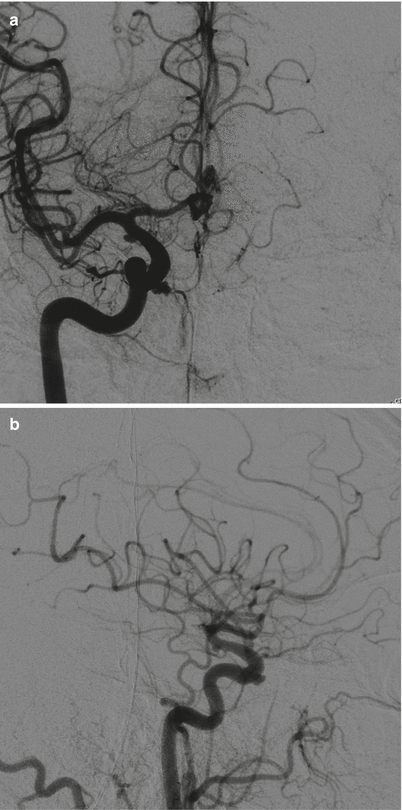
Fig. 74.2
DSA images of pseudoaneurysm. (a) Anterior-posterior projection. (b) Lateral projection following right common carotid artery (CCA) injection. An irregularly shaped, anteriorly/medially projecting aneurysm is seen arising from the cavernous segment of the ICA. An additional small, right supraclinoid aneurysm is also present, likely an incidental posterior communicating artery (PCOM) aneurysm
74.3 Clinical and Surgical Management
Prevention of an ICA injury is of paramount importance for any endoscopic skull base operation. Several key steps can be used to minimize the risk of ICA injury, including optimal patient positioning, microDoppler ultrasonography, neuronavigation, and avoidance of sharp instruments in the region of the ICA [9].
In the event of an intraoperative ICA injury, rapid packing of the site of bleeding is recommended. This often requires the use of optimal suctioning techniques and positioning of the endoscope, in order to identify the site and selectively pack it off. Possible packing materials include gelatin sponge, cottonoids, gauze sponges, or autologous muscle, fat, or fascia. Autologous muscle grafting is the prefered packing/repair material.
It is critical to avoid overpacking the site of an acute arterial bleed, as overpacking may result in worsened avulsion or complete occlusion of the parent vessel.
Close communication with the anesthesia team is mandatory in the event of an ICA injury. When possible, enough time should be given to allow them to catch up and stabilize the patient from a physiological standpoint, with fluids, pressors, blood, and so forth.
Once the patient and the site of arterial injury have been stabilized, the patient is usually transported to the angiography suite for angiography and possible embolization of the pseudoaneurysm, which can be done using various techniques, including the use of coils, balloons, particles, or glue.
In some cases, a patient’s pseudoaneurysm will be temporarily embolized, and a definitive procedure (stent insertion or flow diversion with or without coiling) may be performed later, as such procedures often require concomitant anticoagulation [10].
If the pseudoaneurysm cannot be successfully coiled, a number of endovascular and open surgical alternatives exist. A balloon test occlusion of the ICA may be performed, and the parent artery sacrificed if the patient passes the occlusion test. If occlusion of the ICA is not possible, some form of ECA-ICA bypass may be necessary to provide alternate means of perfusion prior to sacrificing the parent vessel.
Stay updated, free articles. Join our Telegram channel

Full access? Get Clinical Tree


